AUCTORES
Globalize your Research

Research Article | DOI: https://doi.org/10.31579/2768-2757/036
1Division of Vascular and Endovascular Surgery, Mayo Clinic College of Medicine, Rochester, MN, USA.
2Gonda Vascular Center, Mayo Clinic, Rochester, MN, USA.
3INSERM Clinical Investigation Center CIC 1414, Rennes, France.
4Laboratory of Vascular Investigations, University Hospital, Angers, France; and Faculty of Medicine, LUNAM University, Angers, France.
5Department of Biostatistics, Mayo Clinic College of Medicine, Rochester, MN, USA.
*Corresponding Author: Indrani Sen, Division of Vascular and Endovascular Surgery, Mayo Clinic College of Medicine, 200 First Street SW, Rochester, MN 55905, USA.
Citation: I Sen, M Kalra, David A Liedl, G Mahe, P Abraham,et al. (2022). Exercise Transcutaneous Oxygen Pressure (Extcpo2) Measurement is a Reliable Method of Evaluation of Internal Iliac Artery flow Compromise. Journal of Clinical Surgery and Research. 3(2); DOI:10.31579/2768-2757/036
Copyright: ©2022 Indrani Sen, This is an open-access article distributed under the terms of the Creative Commons Attribution License, which permits unrestricted use, distribution, and reproduction in any medium, provided the original author and source are credited.
Received: 27 October 2021 | Accepted: 03 January 2022 | Published: 14 January 2022
Keywords: buttock claudication; peripheral artery disease; exercise test; oximetry; transcutaneous oxygen pressure
Clinical differentiation of buttock claudication from pseudo-claudication (neuromuscular etiology) is often challenging and cannot be diagnosed non-invasively using standard ankle-brachial index (ABI) with exercise. The aim of this study was to evaluate the reliability of exercise transcutaneous oximetry (ExTcPO2) to accurately detect significant internal iliac artery (IIA) inflow stenosis.
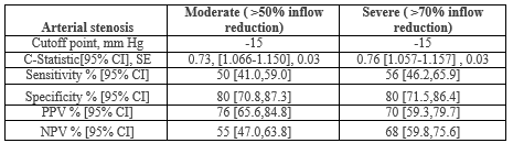
Methods: Data from a prospectively maintained registry of consecutive patients undergoing ExTcPO2 for evaluation of buttock and hip exertional discomfort as well as arterial imaging within 12 months were retrospectively analyzed. ExTcPO2 was performed on a treadmill (10% slope; 2 mph speed); measured at buttocks, upper back and calf (rest, with exercise) to document the baseline normal, degree of change with exercise and recovery patterns. A Delta from Resting Oxygen Pressure (DROP) (buttock- back TcPO2 mmHg) ≥15 mm Hg was considered significant. A blinded physician performed aorto-iliac arterial stenosis quantification and receiver operating characteristic (ROC) curve analysis was used to determine sensitivity and specificity to diagnose severe inflow reduction (≥70%) based upon a diagnostic DROP ≥15 mm Hg.
Results: One hundred and eleven patients (M:F::79:32, mean age 70, range 18-90 years) with available concomitant imaging (CTA 90, DUS 21) were included in the study. Indications for testing were suspected vascular (82), or neuromuscular (29) symptoms. ExTcPO2 study confirmed the clinical suspicion of the state of IAA inflow in 81% (91/111) of patients. DROP ≥15 mm Hg had a sensitivity, specificity, PPV, NPV of 56, 80, 70, and 68% respectively for prediction of severe IIA inflow reduction (p value <0.001; OR 1.116, 95% CI 1.057-1.157; C- statistic 0.76, SE=0.03).
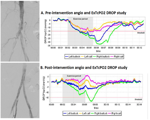
Aorto-iliac arterial reconstruction to treat severe IIA flow compromise was performed in 16 patients. Revascularization was Direct Antegrade in 12 patients (IIA PTA /stenting – 4, aorto-ilac stenting /endarterectomy – 9), Direct Retrograde (aorto-biiliac bypass) in 1 and Indirect via increased collateral flow (profundoplasty) in 3. Post-operatively symptom relief was noted in 15/16 patients and objectively demonstrated on ExTcPo2 in 6/7 (Figure 2). One patient did not improve clinically, or on EXTcPO2 following Indirect revascularization.
Conclusion: Exercise transcutaneous oximetry can reliably diagnose significant IIA inflow stenosis. It is simple, non-invasive and can serve as a valuable screening tool to differentiate buttock claudication from other neuromuscular causes of low back, buttock and hip discomfort as well as post-operatively to confirm adequacy of aorto-iliac revascularization in conjunction with standard, noninvasive vascular evaluation with ABI.
Low back pain associated with hip, buttock, or thigh pain is a common problem in the elderly. Causation is often multifactorial; secondary to peripheral arterial disease (PAD) or other diseases such as lumbar spinal stenosis, sciatica, or hip osteoarthritis. True claudication (pain that is absent at rest, appears during exercise, reproducible and relieved by rest) is easier to differentiate form pseudoclaudicaiton when it occurs in the calf or thigh in the lower limb (distal claudication) [1]. Even so, it necessitates the use of noninvasive physiological assessments. The American College of Cardiology/American Heart Association (ACC/AHA) statements suggest measuring ankle-brachial indices (ABIs), pulse volume recordings, segmental pressures, duplex ultrasound and/or exercising testing with ABI to evaluate claudication versus pseudo-claudication to integrate the clinical and physiologic information [2]. However, these tests are used mainly in the diagnosis and management of distal claudication; these and other tests like Near Infra-Red Spectroscopy (NIRS) and penile-brachial index have not proven reliable in diagnosing proximal claudication as they do not directly assess the arterial beds involved in causation of the latter [3, 4].
Exercise-TcPO2 has been used in France to diagnose proximal claudication due to PAD with a good sensitivity (79%) and specificity (86%) to detect significant lesions (stenosis⩾75%) in the arterial tree of the pelvic circulation when compared to angiography as a gold standard [5]. We adopted a technique of exercise transcutaneous oxygen pressure measurement (exercise-TcPO2) since 2003 and had previously reported the first case in the United States of a patient with proximal claudication being evaluated by an exercise-TcPO2 protocol both before and after angioplasty [6]. In this study we report our results on the role of exercise-TcPO2 in the diagnosis and follow up of patients with proximal claudication due to PAD and the clinical- imaging correlation demonstrating its utility.
Study design and population
Data from a prospectively maintained registry of consecutive patients undergoing ExTcPO2 between January 2013 to January 2017 for evaluation of buttock and hip exertional discomfort as well as arterial imaging within 12 months were retrospectively analyzed. Patients being referred to the lab were those who needed differentiation of vasculogenic from neurogenic claudication and to evaluate the relative severity of buttock vs calf claudication. Demographic characteristics such as age,
gender, nature of symptoms, location and laterality of symptoms, nature of vascular intervention and post procedure outcome were recorded. Procedural details of revascularization were noted. Aorto-iliac arterial reconstruction to treat severe IIA flow compromise was classified as direct antegrade (IIA PTA /stenting; aorto-iliac stenting /femoral endarterectomy), direct retrograde (aorto-biiliac/femoral bypass) and Indirect via increased collateral flow (profundoplasty). The study was approved by the Institutional Review Board.
ExTcPO2 testing protocol
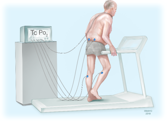
Exercise-TcPO2 was performed according to the published French algorithm using calibrated TcPO2 probes (TCOM/TcPO2;PF 5040 TcPO2/CO2 Unit; Perimed®; Jarfalla, Sweden) using a 5-electrode system. The temperature of each probe was set to 45°C, which allows maximal vasodilation and decreases the arterial to skin surface oxygen pressure gradient. A reference electrode (chest probe) was placed between the scapulae to measure systemic changes in TcPO2 during exercise. One electrode was positioned on each buttock, 4 to 5 cm behind the bony prominence of the trochanter, and on the calf (Figure 2). Once the electrodes were in position, baseline values were obtained with the patient standing for 10 minutes. Exercise was performed on a treadmill at a 10% slope and a speed of up to 2mph. A 12-lead ECG monitored heart rate and rhythm for ischemic changes during the exercise test procedure. The patient was encouraged to walk for the longest time possible to mimic symptoms. Exercise was discontinued at the patient’s request (or, by protocol, up to maximum exercise duration of 12 minutes). The measurements from the TcPO2 electrodes were used to calculate the Delta from Resting Oxygen Pressure (DROP) index (expressed in mmHg), the absolute change in TcPO2 from resting value in each of the three limb probes, corrected for the absolute change in TcPO2 at the chest electrode. The equation for the DROP index is as follows:
DROP(site )= [ PO2 (site)t t - PO2 (site )t 0 ]- [ PO2 (chest)t t - PO2 (chest)t 0 ]
PO2(site)tt is the oxygen pressure at a measurement site at time t, PO2(site)t0 is the mean oxygen pressure at a measurement site over the baseline resting period; PO2(chest)tt is the oxygen pressure at a chest site at time t and PO2(chest)t0 is the mean oxygen pressure at a chest site over the resting period. The DROP index was automatically calculated and displayed graphically by a dedicated software package that allowed real-time monitoring of DROP values at the levels where the probes were placed. The recovery pattern of the TCo2 was also noted. A Delta from Resting Oxygen Pressure (DROP) (buttock- back TcPO2 mmHg) ≥15 mm Hg was considered significant.
Imaging
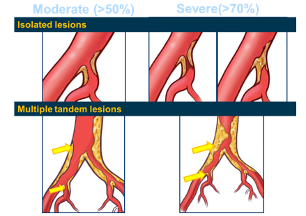
A physician blinded to the results of the ExTcPO2 performed the aorto-iliac arterial stenosis quantification. Imaging characteristics were reviewed to classify aortic, common iliac (CIA), internal iliac (IIA), external iliac (EIA), common femoral (CFA), profunda (PFA) and superficial femoral (SFA) lesions into nil (less than 30%), mild (31-50%), moderate (51-70%) or severe stenosis (greater than 70% stenosis or occlusion). Based on a composite result of the inflow characteristics, a total score was assigned to the degree of inflow compromise at the internal iliac on either limb. Patients with a mild internal iliac stenosis with two tandem proximal CIA or aortic lesions were assigned a higher grade, e.g. if the proximal aortic and CIA lesions were both severe/ occluded, the resultant IIA inflow reduction was graded as severe (Figure 1). In patients with a mild internal iliac stenosis with a single proximal moderate CIA/ aortic lesion the resultant IIA inflow reduction was graded as moderate. In those with isolated internal iliac artery stenosis, the lesion was graded based on the IIA disease alone. In the presence of multiple tandem lesions upstream from the internal iliac artery, the degree of IIA inflow reduction was upgraded. Thus, a composite internal iliac inflow reduction grade: nil (less than 30%), mild (<50>
A physician blinded to the results of Ex-TcPO2 performed aorto-iliac arterial stenosis quantification and receiver operating characteristic (ROC) curve analysis was used to determine sensitivity and specificity to diagnose severe inflow reduction (≥70%) based upon a diagnostic DROP ≥15 mm Hg. Data from patients who underwent revascularization and had a post procedure ExTcPO2 was also analyzed to study its role in outcome assessment. The clinical correlation of the DROP results was noted for the entire study cohort.
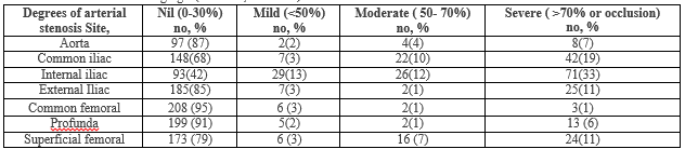
One hundred and eleven patients (M: F:79:32, mean age 70, range 18-90 years) with available concomitant imaging (CTA 90, DUS 21) were included in the study. The Exercise-TcPO2 study was performed for clinical suspicion of vascular claudication in 82 patients (120 limbs) and neuromuscular/ multifactorial symptoms in the remaining 29 patients (42 limbs). The Exercise-TcPO2 study was positive in 56 patients (85 limbs); 45/82 patients with vascular claudication and 11/29 patients with suspected neuromuscular symptoms. ExTcPO2 study confirmed the clinical suspicion of the etiology of the pelvic discomfort being exercise induced reduction of IIA inflow in 81% (91/111) of patients. Arterial imaging included CT angiogram in 90 patients (180limbs) and ultrasound in 21 patients (41 limbs). Patients were classified into mild, moderate or severe depending on the distribution of the stenosis (Table1). Twenty nine patients who did not undergo arterial imaging were excluded from the study.
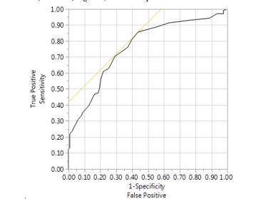
On receiver operating characteristic (ROC) curve analysis DROP ≥15 mm Hg had a sensitivity, specificity, PPV, NPV of 56, 80, 70, and 68% respectively for prediction of severe IIA inflow reduction (p value <0 SE=0.03, SE=0.03)>
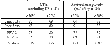
In 27 patients (34 limbs) on imaging studies significant stenosis limiting IIA inflow was noted but ExTcPO2 was negative. The study was limited by severe dyspnea in 2 and lower extremity claudication in the calf/ thigh in 9 that preceded the onset of buttock claudication, symptoms that limited the walking distance and led to early termination of the study before buttock claudication could develop. Subgroup analysis excluding those with ultrasound imaging alone or those patients unable to complete the protocol revealed an improvement in all results (Table 3).
Aorto-iliac arterial reconstruction to treat severe IIA flow compromise was performed in 16 patients. Revascularization was Direct Antegrade in 12 patients (IIA PTA /stenting – 4, aorto-ilac stenting /femoral endarterectomy – 9), Direct Retrograde (aorto-bifemoral bypass) in 1 and Indirect via increased collateral flow (profundoplasty) in 3. Post-operatively symptom relief was noted in 15/16 patients and objectively demonstrated on ExTcPo2 in 6/7. This was useful for its ability to evaluate comparative severity of calf vs buttock claudication (Figure 4). Similar to standard non invasive testing, it can be used post-operatively to confirm adequacy of aorto-iliac revascularization (Figure 5). One patient did not improve clinically, or on ExTcPO2 following Indirect retrograde revascularization. This patient underwent profundoplasty for disabling thigh and buttock claudication (imaging had revealed long segment occlusion of the IIA with poor distal reformation and occlusion of the profunda). Post procedure, he had improvement of the thigh claudication but no improvement in the buttock claudication which could be explained by the poor improvement in collateral flow secondary to the long segment IIA occlusion.

The prevalence of proximal vascular claudication or buttock claudication has come to light in the post EVAR era when internal iliac artery sacrifice was performed frequently. Similar symptoms can be caused by various orthopaedic disorders and a combination of these frequently exist in elderly patients. Clinical differentiation of buttock claudication from pseudo-claudication (neuromuscular etiology) is often challenging. Patients are typically older and present with atypical lower back, hip, buttock, or thigh pain arising from common iliac and/or internal iliac flow limiting lesions. Many patients present with atypical symptoms not typical of claudication or with concomitant neuromuscular and orthopedic conditions that may also give rise to pain (pseudo-claudication) indistinguishable form that produced by arterial disease [1]. It is usually asymptomatic, as worsening in claudication may be masked by a compensatory decline in functional performance in spite of disease progression [7]. Evaluation of proximal claudication is challenging. Standard noninvasive vascular tests measure axial lower extremity but not direct internal iliac or abdominal-pelvic collateral flow.Visualization of the internal iliac arteries using duplex may be difficult secondary to its location, and inflow abnormalities cannot be diagnosed non-invasively using standard tests like ankle-brachial index (ABI) with exercise that are designed to test axial blood flow. Transcutaneous oxygen tension measurement reflects the metabolic state of target tissues and exercise transcutaneous oxygen pressure (Exercise-TcPO2) has previously been reported to be useful in the diagnosis of proximal claudication due to PAD [6]. This study demonstrates the utility of ExTco2 in diagnosing the role of internal iliac artery flow compromise as the significant contributing cause of proximal walking induced pain, even while multiple potential other etiologies are present.
Patients often present with concomitant lower limb claudication and conventional non-invasive testing with ABI/ exercise ABI/Toe pressure/ Pulse volume recordings remain first line. Non-invasive testing using transcutaneous oxygen tension measurements in the lower limb has a better sensitivity/specificity in diagnosing severe flow limiting lesions, its role in diagnosing moderate or mild lesions is thought to be poor. This may not be a limitation in the internal iliac bed; as intervention is most often offered only for those with critical lesions. Also, the gluteal skin and underlying muscle beds have a common supply from the internal iliac and the calculation of the DROP index helps in cancelling out the effect of an exercise induced drop in skin Tco2 purely due to shunting of blood by dilatation of intramuscular vessels similar to what is seen in the pedal circulation.
In such patients with both proximal (buttock) and distal (axial) vascular compromise the use of a 5-electrode system also allows for 4 different sites can be evaluated in the same time. This also allows for calculation of a proximal and distal DROP; those with a proximal DROP that is higher than the distal are more likely to have an internal iliac artery flow limiting proximal lesion [8]. As demonstrated by our study, Ex-TcPO2 with calculation of a DROP index allows for functional assessment of the internal iliac artery and distal lower extremity circulation during walking and helps determine the area with the greater arterial inflow compromise that is responsible for limiting ambulation, helping guide the location and extent of revascularization that is most likely to provide symptom relief.
In the final analysis identification of internal iliac artery flow compromise with ExTcPo2 is especially relevant for three reasons: first, it allows for the assessment of blood flow during exercise; which is a more accurate assessment of the underlying physiology as during exercise is when symptoms occur, helps determine the maximal walking distance and reveals other causes of exercise limitation. This is particularly useful in patients with a combination of etiologies that limit ambulation; Ex-TCPo2 helps determine the more severe etiology thereby guiding evaluation and treatment. Second, it also allows for assessment of the arterial physiology in those with calcified or non-compressible vessels (diabetes, renal insufficiency, advanced age) where ABPI may be falsely elevated. Ex-TcPo2 is accurate to detect IIA flow compromise ≥70% that is similar to ultrasound to detect aorto-iliac stenoses. [9, 10] We thus included the imaging results of those with ultrasound imaging alone as this is a more realistic clinical scenario in this patient cohort. Third, the degree of DROP in each area helps guide the site and extent of vascular intervention that the patient may benefit from.
Ex-TcPO2 has been validated against angiography in two previous studies [5, 11] and CT-angiogram in one study [8] with a good sensitivity/specificity of this method to detect proximal arterial stenosis. This is the first report of validation of this technique in the United States. Abraham et.al reported on the utility of Ex-TcPo2 with a cutoff point of -15 mm Hg DROP demonstrating a 79%/86% sensitivity/specificity in the diagnosis of internal iliac flow compromise; but they did not grade the degree or laterality of the internal iliac stenosis [5]. The study by Koch et. al included 34 patients with suspected claudication and noted that exercise-TcPo2 using a proximal minimal DROP value ≤−15 mm Hg or a distal minimal DROP value ≤−16 mm Hg is accurate to diagnose arterial stenosis especially stenosis ≥60% on the lower limbs. However this study, stenosis was assessed at a single level (aortic/ CIA/IIA) was used for analysis and did not analyze the effect of a combined aortic/ common iliac lesion. We used an algorithm to account for tandem lesions and tried to correlate the potential physiologic significance of lesions by correlating it to ex-TcPo2. Koch et.al reported a specificity of 76% for a high grade (>70%) stenosis which is similar to our results. Our sensitivity is lower, this may be because the study was negative in those patients who developed early calf/ thigh claudication or dyspnea that limited the walking distance and led to early termination of the study. The high specificity of the study is similar to that reported by other authors and supports the utility of Ex-TcPO2 in diagnosing proximal claudication.
We demonstrate that ExTCo2 improves after treatment and is useful in assessing outcome, follow up and surveillance following these procedures. The importance of preserving the pelvic circulation to prevent gluteal claudication is well recognized [12]. Both open and endovascular intervention may be necessary for symptom relief; with a good 5 year patency and freedom from buttock claudication (both around 80%) [12-15]. In addition, an improvement in the ExTCo2 following intervention in a patient with persistent symptoms of hip pain should prompt more aggressive evaluation and management of non-vascular etiologies.
Our study has some limitations, the overall numbers remain small and we have not analyzed the effect of the pretest probability based on the nature of symptoms and its correlation with internal iliac disease based on imaging. It is time consuming, automation is essential, current software is proprietary. However, this does not affect its obvious utility in diagnosis or follow up as those without a higher pretest probability are not likely to be advised this test due to its niche utility. Also, a drift correction to correct a potential drift of the O2 pressure measurement with time at each electrode was not used in all patients.
Exercise transcutaneous oximetry can reliably diagnose significant IIA inflow stenosis. It is a simple, non-invasive and can serve as a valuable screening tool to differentiate buttock claudication from other neuromuscular causes of low back, buttock and hip discomfort as well as the ability to evaluate comparative severity of calf vs buttock claudication. Similar to standard noninvasive testing, it can be used post-operatively to confirm adequacy of aorto-iliac revascularization and for surveillance.
IS, DL: Data collection, analysis, statistics, drafting manuscript
WH: PW: Analysis, statistics, drafting manuscript
GM, PA, MK: Concept, data collection, analysis, statistics, drafting manuscript, final approval
Clearly Auctoresonline and particularly Psychology and Mental Health Care Journal is dedicated to improving health care services for individuals and populations. The editorial boards' ability to efficiently recognize and share the global importance of health literacy with a variety of stakeholders. Auctoresonline publishing platform can be used to facilitate of optimal client-based services and should be added to health care professionals' repertoire of evidence-based health care resources.

Journal of Clinical Cardiology and Cardiovascular Intervention The submission and review process was adequate. However I think that the publication total value should have been enlightened in early fases. Thank you for all.

Journal of Women Health Care and Issues By the present mail, I want to say thank to you and tour colleagues for facilitating my published article. Specially thank you for the peer review process, support from the editorial office. I appreciate positively the quality of your journal.

Journal of Clinical Research and Reports I would be very delighted to submit my testimonial regarding the reviewer board and the editorial office. The reviewer board were accurate and helpful regarding any modifications for my manuscript. And the editorial office were very helpful and supportive in contacting and monitoring with any update and offering help. It was my pleasure to contribute with your promising Journal and I am looking forward for more collaboration.

We would like to thank the Journal of Thoracic Disease and Cardiothoracic Surgery because of the services they provided us for our articles. The peer-review process was done in a very excellent time manner, and the opinions of the reviewers helped us to improve our manuscript further. The editorial office had an outstanding correspondence with us and guided us in many ways. During a hard time of the pandemic that is affecting every one of us tremendously, the editorial office helped us make everything easier for publishing scientific work. Hope for a more scientific relationship with your Journal.

The peer-review process which consisted high quality queries on the paper. I did answer six reviewers’ questions and comments before the paper was accepted. The support from the editorial office is excellent.

Journal of Neuroscience and Neurological Surgery. I had the experience of publishing a research article recently. The whole process was simple from submission to publication. The reviewers made specific and valuable recommendations and corrections that improved the quality of my publication. I strongly recommend this Journal.

Dr. Katarzyna Byczkowska My testimonial covering: "The peer review process is quick and effective. The support from the editorial office is very professional and friendly. Quality of the Clinical Cardiology and Cardiovascular Interventions is scientific and publishes ground-breaking research on cardiology that is useful for other professionals in the field.

Thank you most sincerely, with regard to the support you have given in relation to the reviewing process and the processing of my article entitled "Large Cell Neuroendocrine Carcinoma of The Prostate Gland: A Review and Update" for publication in your esteemed Journal, Journal of Cancer Research and Cellular Therapeutics". The editorial team has been very supportive.

Testimony of Journal of Clinical Otorhinolaryngology: work with your Reviews has been a educational and constructive experience. The editorial office were very helpful and supportive. It was a pleasure to contribute to your Journal.

Dr. Bernard Terkimbi Utoo, I am happy to publish my scientific work in Journal of Women Health Care and Issues (JWHCI). The manuscript submission was seamless and peer review process was top notch. I was amazed that 4 reviewers worked on the manuscript which made it a highly technical, standard and excellent quality paper. I appreciate the format and consideration for the APC as well as the speed of publication. It is my pleasure to continue with this scientific relationship with the esteem JWHCI.

This is an acknowledgment for peer reviewers, editorial board of Journal of Clinical Research and Reports. They show a lot of consideration for us as publishers for our research article “Evaluation of the different factors associated with side effects of COVID-19 vaccination on medical students, Mutah university, Al-Karak, Jordan”, in a very professional and easy way. This journal is one of outstanding medical journal.

Dear Hao Jiang, to Journal of Nutrition and Food Processing We greatly appreciate the efficient, professional and rapid processing of our paper by your team. If there is anything else we should do, please do not hesitate to let us know. On behalf of my co-authors, we would like to express our great appreciation to editor and reviewers.

As an author who has recently published in the journal "Brain and Neurological Disorders". I am delighted to provide a testimonial on the peer review process, editorial office support, and the overall quality of the journal. The peer review process at Brain and Neurological Disorders is rigorous and meticulous, ensuring that only high-quality, evidence-based research is published. The reviewers are experts in their fields, and their comments and suggestions were constructive and helped improve the quality of my manuscript. The review process was timely and efficient, with clear communication from the editorial office at each stage. The support from the editorial office was exceptional throughout the entire process. The editorial staff was responsive, professional, and always willing to help. They provided valuable guidance on formatting, structure, and ethical considerations, making the submission process seamless. Moreover, they kept me informed about the status of my manuscript and provided timely updates, which made the process less stressful. The journal Brain and Neurological Disorders is of the highest quality, with a strong focus on publishing cutting-edge research in the field of neurology. The articles published in this journal are well-researched, rigorously peer-reviewed, and written by experts in the field. The journal maintains high standards, ensuring that readers are provided with the most up-to-date and reliable information on brain and neurological disorders. In conclusion, I had a wonderful experience publishing in Brain and Neurological Disorders. The peer review process was thorough, the editorial office provided exceptional support, and the journal's quality is second to none. I would highly recommend this journal to any researcher working in the field of neurology and brain disorders.
Dear Agrippa Hilda, Journal of Neuroscience and Neurological Surgery, Editorial Coordinator, I trust this message finds you well. I want to extend my appreciation for considering my article for publication in your esteemed journal. I am pleased to provide a testimonial regarding the peer review process and the support received from your editorial office. The peer review process for my paper was carried out in a highly professional and thorough manner. The feedback and comments provided by the authors were constructive and very useful in improving the quality of the manuscript. This rigorous assessment process undoubtedly contributes to the high standards maintained by your journal.

International Journal of Clinical Case Reports and Reviews. I strongly recommend to consider submitting your work to this high-quality journal. The support and availability of the Editorial staff is outstanding and the review process was both efficient and rigorous.

Thank you very much for publishing my Research Article titled “Comparing Treatment Outcome Of Allergic Rhinitis Patients After Using Fluticasone Nasal Spray And Nasal Douching" in the Journal of Clinical Otorhinolaryngology. As Medical Professionals we are immensely benefited from study of various informative Articles and Papers published in this high quality Journal. I look forward to enriching my knowledge by regular study of the Journal and contribute my future work in the field of ENT through the Journal for use by the medical fraternity. The support from the Editorial office was excellent and very prompt. I also welcome the comments received from the readers of my Research Article.

Dear Erica Kelsey, Editorial Coordinator of Cancer Research and Cellular Therapeutics Our team is very satisfied with the processing of our paper by your journal. That was fast, efficient, rigorous, but without unnecessary complications. We appreciated the very short time between the submission of the paper and its publication on line on your site.

I am very glad to say that the peer review process is very successful and fast and support from the Editorial Office. Therefore, I would like to continue our scientific relationship for a long time. And I especially thank you for your kindly attention towards my article. Have a good day!

"We recently published an article entitled “Influence of beta-Cyclodextrins upon the Degradation of Carbofuran Derivatives under Alkaline Conditions" in the Journal of “Pesticides and Biofertilizers” to show that the cyclodextrins protect the carbamates increasing their half-life time in the presence of basic conditions This will be very helpful to understand carbofuran behaviour in the analytical, agro-environmental and food areas. We greatly appreciated the interaction with the editor and the editorial team; we were particularly well accompanied during the course of the revision process, since all various steps towards publication were short and without delay".

I would like to express my gratitude towards you process of article review and submission. I found this to be very fair and expedient. Your follow up has been excellent. I have many publications in national and international journal and your process has been one of the best so far. Keep up the great work.

We are grateful for this opportunity to provide a glowing recommendation to the Journal of Psychiatry and Psychotherapy. We found that the editorial team were very supportive, helpful, kept us abreast of timelines and over all very professional in nature. The peer review process was rigorous, efficient and constructive that really enhanced our article submission. The experience with this journal remains one of our best ever and we look forward to providing future submissions in the near future.

I am very pleased to serve as EBM of the journal, I hope many years of my experience in stem cells can help the journal from one way or another. As we know, stem cells hold great potential for regenerative medicine, which are mostly used to promote the repair response of diseased, dysfunctional or injured tissue using stem cells or their derivatives. I think Stem Cell Research and Therapeutics International is a great platform to publish and share the understanding towards the biology and translational or clinical application of stem cells.

I would like to give my testimony in the support I have got by the peer review process and to support the editorial office where they were of asset to support young author like me to be encouraged to publish their work in your respected journal and globalize and share knowledge across the globe. I really give my great gratitude to your journal and the peer review including the editorial office.

I am delighted to publish our manuscript entitled "A Perspective on Cocaine Induced Stroke - Its Mechanisms and Management" in the Journal of Neuroscience and Neurological Surgery. The peer review process, support from the editorial office, and quality of the journal are excellent. The manuscripts published are of high quality and of excellent scientific value. I recommend this journal very much to colleagues.

Dr.Tania Muñoz, My experience as researcher and author of a review article in The Journal Clinical Cardiology and Interventions has been very enriching and stimulating. The editorial team is excellent, performs its work with absolute responsibility and delivery. They are proactive, dynamic and receptive to all proposals. Supporting at all times the vast universe of authors who choose them as an option for publication. The team of review specialists, members of the editorial board, are brilliant professionals, with remarkable performance in medical research and scientific methodology. Together they form a frontline team that consolidates the JCCI as a magnificent option for the publication and review of high-level medical articles and broad collective interest. I am honored to be able to share my review article and open to receive all your comments.

“The peer review process of JPMHC is quick and effective. Authors are benefited by good and professional reviewers with huge experience in the field of psychology and mental health. The support from the editorial office is very professional. People to contact to are friendly and happy to help and assist any query authors might have. Quality of the Journal is scientific and publishes ground-breaking research on mental health that is useful for other professionals in the field”.

Dear editorial department: On behalf of our team, I hereby certify the reliability and superiority of the International Journal of Clinical Case Reports and Reviews in the peer review process, editorial support, and journal quality. Firstly, the peer review process of the International Journal of Clinical Case Reports and Reviews is rigorous, fair, transparent, fast, and of high quality. The editorial department invites experts from relevant fields as anonymous reviewers to review all submitted manuscripts. These experts have rich academic backgrounds and experience, and can accurately evaluate the academic quality, originality, and suitability of manuscripts. The editorial department is committed to ensuring the rigor of the peer review process, while also making every effort to ensure a fast review cycle to meet the needs of authors and the academic community. Secondly, the editorial team of the International Journal of Clinical Case Reports and Reviews is composed of a group of senior scholars and professionals with rich experience and professional knowledge in related fields. The editorial department is committed to assisting authors in improving their manuscripts, ensuring their academic accuracy, clarity, and completeness. Editors actively collaborate with authors, providing useful suggestions and feedback to promote the improvement and development of the manuscript. We believe that the support of the editorial department is one of the key factors in ensuring the quality of the journal. Finally, the International Journal of Clinical Case Reports and Reviews is renowned for its high- quality articles and strict academic standards. The editorial department is committed to publishing innovative and academically valuable research results to promote the development and progress of related fields. The International Journal of Clinical Case Reports and Reviews is reasonably priced and ensures excellent service and quality ratio, allowing authors to obtain high-level academic publishing opportunities in an affordable manner. I hereby solemnly declare that the International Journal of Clinical Case Reports and Reviews has a high level of credibility and superiority in terms of peer review process, editorial support, reasonable fees, and journal quality. Sincerely, Rui Tao.

Clinical Cardiology and Cardiovascular Interventions I testity the covering of the peer review process, support from the editorial office, and quality of the journal.

Clinical Cardiology and Cardiovascular Interventions, we deeply appreciate the interest shown in our work and its publication. It has been a true pleasure to collaborate with you. The peer review process, as well as the support provided by the editorial office, have been exceptional, and the quality of the journal is very high, which was a determining factor in our decision to publish with you.

The peer reviewers process is quick and effective, the supports from editorial office is excellent, the quality of journal is high. I would like to collabroate with Internatioanl journal of Clinical Case Reports and Reviews journal clinically in the future time.

Clinical Cardiology and Cardiovascular Interventions, I would like to express my sincerest gratitude for the trust placed in our team for the publication in your journal. It has been a true pleasure to collaborate with you on this project. I am pleased to inform you that both the peer review process and the attention from the editorial coordination have been excellent. Your team has worked with dedication and professionalism to ensure that your publication meets the highest standards of quality. We are confident that this collaboration will result in mutual success, and we are eager to see the fruits of this shared effort.

Dear Dr. Jessica Magne, Editorial Coordinator 0f Clinical Cardiology and Cardiovascular Interventions, I hope this message finds you well. I want to express my utmost gratitude for your excellent work and for the dedication and speed in the publication process of my article titled "Navigating Innovation: Qualitative Insights on Using Technology for Health Education in Acute Coronary Syndrome Patients." I am very satisfied with the peer review process, the support from the editorial office, and the quality of the journal. I hope we can maintain our scientific relationship in the long term.

Dear Monica Gissare, - Editorial Coordinator of Nutrition and Food Processing. ¨My testimony with you is truly professional, with a positive response regarding the follow-up of the article and its review, you took into account my qualities and the importance of the topic¨.

Dear Dr. Jessica Magne, Editorial Coordinator 0f Clinical Cardiology and Cardiovascular Interventions, The review process for the article “The Handling of Anti-aggregants and Anticoagulants in the Oncologic Heart Patient Submitted to Surgery” was extremely rigorous and detailed. From the initial submission to the final acceptance, the editorial team at the “Journal of Clinical Cardiology and Cardiovascular Interventions” demonstrated a high level of professionalism and dedication. The reviewers provided constructive and detailed feedback, which was essential for improving the quality of our work. Communication was always clear and efficient, ensuring that all our questions were promptly addressed. The quality of the “Journal of Clinical Cardiology and Cardiovascular Interventions” is undeniable. It is a peer-reviewed, open-access publication dedicated exclusively to disseminating high-quality research in the field of clinical cardiology and cardiovascular interventions. The journal's impact factor is currently under evaluation, and it is indexed in reputable databases, which further reinforces its credibility and relevance in the scientific field. I highly recommend this journal to researchers looking for a reputable platform to publish their studies.

Dear Editorial Coordinator of the Journal of Nutrition and Food Processing! "I would like to thank the Journal of Nutrition and Food Processing for including and publishing my article. The peer review process was very quick, movement and precise. The Editorial Board has done an extremely conscientious job with much help, valuable comments and advices. I find the journal very valuable from a professional point of view, thank you very much for allowing me to be part of it and I would like to participate in the future!”

Dealing with The Journal of Neurology and Neurological Surgery was very smooth and comprehensive. The office staff took time to address my needs and the response from editors and the office was prompt and fair. I certainly hope to publish with this journal again.Their professionalism is apparent and more than satisfactory. Susan Weiner

My Testimonial Covering as fellowing: Lin-Show Chin. The peer reviewers process is quick and effective, the supports from editorial office is excellent, the quality of journal is high. I would like to collabroate with Internatioanl journal of Clinical Case Reports and Reviews.

My experience publishing in Psychology and Mental Health Care was exceptional. The peer review process was rigorous and constructive, with reviewers providing valuable insights that helped enhance the quality of our work. The editorial team was highly supportive and responsive, making the submission process smooth and efficient. The journal's commitment to high standards and academic rigor makes it a respected platform for quality research. I am grateful for the opportunity to publish in such a reputable journal.

My experience publishing in International Journal of Clinical Case Reports and Reviews was exceptional. I Come forth to Provide a Testimonial Covering the Peer Review Process and the editorial office for the Professional and Impartial Evaluation of the Manuscript.

I would like to offer my testimony in the support. I have received through the peer review process and support the editorial office where they are to support young authors like me, encourage them to publish their work in your esteemed journals, and globalize and share knowledge globally. I really appreciate your journal, peer review, and editorial office.

Dear Agrippa Hilda- Editorial Coordinator of Journal of Neuroscience and Neurological Surgery, "The peer review process was very quick and of high quality, which can also be seen in the articles in the journal. The collaboration with the editorial office was very good."

I would like to express my sincere gratitude for the support and efficiency provided by the editorial office throughout the publication process of my article, “Delayed Vulvar Metastases from Rectal Carcinoma: A Case Report.” I greatly appreciate the assistance and guidance I received from your team, which made the entire process smooth and efficient. The peer review process was thorough and constructive, contributing to the overall quality of the final article. I am very grateful for the high level of professionalism and commitment shown by the editorial staff, and I look forward to maintaining a long-term collaboration with the International Journal of Clinical Case Reports and Reviews.

To Dear Erin Aust, I would like to express my heartfelt appreciation for the opportunity to have my work published in this esteemed journal. The entire publication process was smooth and well-organized, and I am extremely satisfied with the final result. The Editorial Team demonstrated the utmost professionalism, providing prompt and insightful feedback throughout the review process. Their clear communication and constructive suggestions were invaluable in enhancing my manuscript, and their meticulous attention to detail and dedication to quality are truly commendable. Additionally, the support from the Editorial Office was exceptional. From the initial submission to the final publication, I was guided through every step of the process with great care and professionalism. The team's responsiveness and assistance made the entire experience both easy and stress-free. I am also deeply impressed by the quality and reputation of the journal. It is an honor to have my research featured in such a respected publication, and I am confident that it will make a meaningful contribution to the field.

"I am grateful for the opportunity of contributing to [International Journal of Clinical Case Reports and Reviews] and for the rigorous review process that enhances the quality of research published in your esteemed journal. I sincerely appreciate the time and effort of your team who have dedicatedly helped me in improvising changes and modifying my manuscript. The insightful comments and constructive feedback provided have been invaluable in refining and strengthening my work".

I thank the ‘Journal of Clinical Research and Reports’ for accepting this article for publication. This is a rigorously peer reviewed journal which is on all major global scientific data bases. I note the review process was prompt, thorough and professionally critical. It gave us an insight into a number of important scientific/statistical issues. The review prompted us to review the relevant literature again and look at the limitations of the study. The peer reviewers were open, clear in the instructions and the editorial team was very prompt in their communication. This journal certainly publishes quality research articles. I would recommend the journal for any future publications.

Dear Jessica Magne, with gratitude for the joint work. Fast process of receiving and processing the submitted scientific materials in “Clinical Cardiology and Cardiovascular Interventions”. High level of competence of the editors with clear and correct recommendations and ideas for enriching the article.

We found the peer review process quick and positive in its input. The support from the editorial officer has been very agile, always with the intention of improving the article and taking into account our subsequent corrections.

My article, titled 'No Way Out of the Smartphone Epidemic Without Considering the Insights of Brain Research,' has been republished in the International Journal of Clinical Case Reports and Reviews. The review process was seamless and professional, with the editors being both friendly and supportive. I am deeply grateful for their efforts.

To Dear Erin Aust – Editorial Coordinator of Journal of General Medicine and Clinical Practice! I declare that I am absolutely satisfied with your work carried out with great competence in following the manuscript during the various stages from its receipt, during the revision process to the final acceptance for publication. Thank Prof. Elvira Farina

Dear Jessica, and the super professional team of the ‘Clinical Cardiology and Cardiovascular Interventions’ I am sincerely grateful to the coordinated work of the journal team for the no problem with the submission of my manuscript: “Cardiometabolic Disorders in A Pregnant Woman with Severe Preeclampsia on the Background of Morbid Obesity (Case Report).” The review process by 5 experts was fast, and the comments were professional, which made it more specific and academic, and the process of publication and presentation of the article was excellent. I recommend that my colleagues publish articles in this journal, and I am interested in further scientific cooperation. Sincerely and best wishes, Dr. Oleg Golyanovskiy.

Dear Ashley Rosa, Editorial Coordinator of the journal - Psychology and Mental Health Care. " The process of obtaining publication of my article in the Psychology and Mental Health Journal was positive in all areas. The peer review process resulted in a number of valuable comments, the editorial process was collaborative and timely, and the quality of this journal has been quickly noticed, resulting in alternative journals contacting me to publish with them." Warm regards, Susan Anne Smith, PhD. Australian Breastfeeding Association.

Dear Jessica Magne, Editorial Coordinator, Clinical Cardiology and Cardiovascular Interventions, Auctores Publishing LLC. I appreciate the journal (JCCI) editorial office support, the entire team leads were always ready to help, not only on technical front but also on thorough process. Also, I should thank dear reviewers’ attention to detail and creative approach to teach me and bring new insights by their comments. Surely, more discussions and introduction of other hemodynamic devices would provide better prevention and management of shock states. Your efforts and dedication in presenting educational materials in this journal are commendable. Best wishes from, Farahnaz Fallahian.

Dear Maria Emerson, Editorial Coordinator, International Journal of Clinical Case Reports and Reviews, Auctores Publishing LLC. I am delighted to have published our manuscript, "Acute Colonic Pseudo-Obstruction (ACPO): A rare but serious complication following caesarean section." I want to thank the editorial team, especially Maria Emerson, for their prompt review of the manuscript, quick responses to queries, and overall support. Yours sincerely Dr. Victor Olagundoye.

Dear Ashley Rosa, Editorial Coordinator, International Journal of Clinical Case Reports and Reviews. Many thanks for publishing this manuscript after I lost confidence the editors were most helpful, more than other journals Best wishes from, Susan Anne Smith, PhD. Australian Breastfeeding Association.

Dear Agrippa Hilda, Editorial Coordinator, Journal of Neuroscience and Neurological Surgery. The entire process including article submission, review, revision, and publication was extremely easy. The journal editor was prompt and helpful, and the reviewers contributed to the quality of the paper. Thank you so much! Eric Nussbaum, MD

Dr Hala Al Shaikh This is to acknowledge that the peer review process for the article ’ A Novel Gnrh1 Gene Mutation in Four Omani Male Siblings, Presentation and Management ’ sent to the International Journal of Clinical Case Reports and Reviews was quick and smooth. The editorial office was prompt with easy communication.

Dear Erin Aust, Editorial Coordinator, Journal of General Medicine and Clinical Practice. We are pleased to share our experience with the “Journal of General Medicine and Clinical Practice”, following the successful publication of our article. The peer review process was thorough and constructive, helping to improve the clarity and quality of the manuscript. We are especially thankful to Ms. Erin Aust, the Editorial Coordinator, for her prompt communication and continuous support throughout the process. Her professionalism ensured a smooth and efficient publication experience. The journal upholds high editorial standards, and we highly recommend it to fellow researchers seeking a credible platform for their work. Best wishes By, Dr. Rakhi Mishra.

Dear Jessica Magne, Editorial Coordinator, Clinical Cardiology and Cardiovascular Interventions, Auctores Publishing LLC. The peer review process of the journal of Clinical Cardiology and Cardiovascular Interventions was excellent and fast, as was the support of the editorial office and the quality of the journal. Kind regards Walter F. Riesen Prof. Dr. Dr. h.c. Walter F. Riesen.

Dear Ashley Rosa, Editorial Coordinator, International Journal of Clinical Case Reports and Reviews, Auctores Publishing LLC. Thank you for publishing our article, Exploring Clozapine's Efficacy in Managing Aggression: A Multiple Single-Case Study in Forensic Psychiatry in the international journal of clinical case reports and reviews. We found the peer review process very professional and efficient. The comments were constructive, and the whole process was efficient. On behalf of the co-authors, I would like to thank you for publishing this article. With regards, Dr. Jelle R. Lettinga.

Dear Clarissa Eric, Editorial Coordinator, Journal of Clinical Case Reports and Studies, I would like to express my deep admiration for the exceptional professionalism demonstrated by your journal. I am thoroughly impressed by the speed of the editorial process, the substantive and insightful reviews, and the meticulous preparation of the manuscript for publication. Additionally, I greatly appreciate the courteous and immediate responses from your editorial office to all my inquiries. Best Regards, Dariusz Ziora

Dear Chrystine Mejia, Editorial Coordinator, Journal of Neurodegeneration and Neurorehabilitation, Auctores Publishing LLC, We would like to thank the editorial team for the smooth and high-quality communication leading up to the publication of our article in the Journal of Neurodegeneration and Neurorehabilitation. The reviewers have extensive knowledge in the field, and their relevant questions helped to add value to our publication. Kind regards, Dr. Ravi Shrivastava.

Dear Clarissa Eric, Editorial Coordinator, Journal of Clinical Case Reports and Studies, Auctores Publishing LLC, USA Office: +1-(302)-520-2644. I would like to express my sincere appreciation for the efficient and professional handling of my case report by the ‘Journal of Clinical Case Reports and Studies’. The peer review process was not only fast but also highly constructive—the reviewers’ comments were clear, relevant, and greatly helped me improve the quality and clarity of my manuscript. I also received excellent support from the editorial office throughout the process. Communication was smooth and timely, and I felt well guided at every stage, from submission to publication. The overall quality and rigor of the journal are truly commendable. I am pleased to have published my work with Journal of Clinical Case Reports and Studies, and I look forward to future opportunities for collaboration. Sincerely, Aline Tollet, UCLouvain.

Dear Ms. Mayra Duenas, Editorial Coordinator, International Journal of Clinical Case Reports and Reviews. “The International Journal of Clinical Case Reports and Reviews represented the “ideal house” to share with the research community a first experience with the use of the Simeox device for speech rehabilitation. High scientific reputation and attractive website communication were first determinants for the selection of this Journal, and the following submission process exceeded expectations: fast but highly professional peer review, great support by the editorial office, elegant graphic layout. Exactly what a dynamic research team - also composed by allied professionals - needs!" From, Chiara Beccaluva, PT - Italy.

Dear Maria Emerson, Editorial Coordinator, we have deeply appreciated the professionalism demonstrated by the International Journal of Clinical Case Reports and Reviews. The reviewers have extensive knowledge of our field and have been very efficient and fast in supporting the process. I am really looking forward to further collaboration. Thanks. Best regards, Dr. Claudio Ligresti

Dear Chrystine Mejia, Editorial Coordinator, Journal of Neurodegeneration and Neurorehabilitation. “The peer review process was efficient and constructive, and the editorial office provided excellent communication and support throughout. The journal ensures scientific rigor and high editorial standards, while also offering a smooth and timely publication process. We sincerely appreciate the work of the editorial team in facilitating the dissemination of innovative approaches such as the Bonori Method.” Best regards, Dr. Matteo Bonori.
