AUCTORES
Globalize your Research

Research Article | DOI: https://doi.org/10.31579/2643-6612/029
1 Department of Oral and Maxillofacial Surgery, Leiden University Medical Center, Leiden, the Netherlands.
2 Mathematical Institute, Leiden University, Niels Bohrweg 1, 2333 CA Leiden, the Netherlands.
3 Medical Statistics, Department of Biomedical Data Sciences, Leiden University Medical Center, Leiden, the Netherlands.
4 Department of Oral and Maxillofacial Surgery, Amphia Hospital, Breda, the Netherlands.
*Corresponding Author: JP Richard van Merkesteyn, Department of Oral and Maxillofacial Surgery, Leiden University Medical Center, Leiden, the Netherlands.
Citation: Jeroen G van Rijsse, Jop P Verweij, M Fiocco, G Mensink, J P Richard van Merkesteyn. (2022). Visible Soft Tissue Contour Change of the Mandible Associated with Osseous Inferior Mandibular Border Defects after BSSO. Dentistry and Oral Maxillofacial Surgery. 5(2); DOI: 10.31579/2643-6612/029
Copyright: © 2022 JP Richard van Merkesteyn, this is an open-access article distributed under the terms of the Creative Commons Attribution License, which permits unrestricted use, distribution, and reproduction in any medium, provided the original author and source are credited.
Received: 16 December 2021 | Accepted: 02 February 2022 | Published: 18 February 2022
Keywords: sagittal split; osteotomy; orthognathic; complication; inferior border
Purpose: Visible soft tissue contour changes of the mandibular inferior border can cause an unaesthetic outcome after bilateral sagittal split osteotomy (BSSO). In some cases, even secondary reconstruction of the mandibular inferior border is needed. The aim of this study was to determine the percentage of unwanted visible contour changes of the inferior mandibular border after BSSO. The impact of potential risk factors for the outcome of interest were also assessed.
Methods: In this retrospective study, consecutive patients who underwent mandibular advancement through BSSO were included. The primary outcome parameter was the presence/absence of a visible contour change at the inferior border of the mandible one year after BSSO. Risk factors of interest included the presence of a radiographic osseous inferior border defect, the amount of mandibular movement, rotation of the occlusal plane, postoperative proximal segment position, pattern of lingual fracture, occurrence of bad split, and presence of third molars during BSSO.
Results: The study sample consisted of 147 patients with a mean follow-up of 13.2 months. A visible contour change was present in 2% of patients (1% of sagittal splits). No secondary reconstructive procedures were performed. A bony defect (osseous inferior border defect) was present in 7% of the sagittal splits. There was a significant association between a visible contour change of the mandibular border and an osseous inferior border defect (p <0.001). None of the other risk factors in this study showed a significant association with soft tissue contour changes.
Conclusion: Visible contour changes of the inferior border of the mandible are a rare complication after BSSO. Osseous inferior border defects of the mandible are a significant risk factor for soft tissue changes.
Bilateral sagittal split osteotomy (BSSO) for mandibular advancement is a procedure that is widely used to treat patients with a class II malocclusion. Well-known complications are bad splits, damage to the inferior alveolar nerve and postoperative infection. (Jop P Verweij et al., 2016). A much less known complication after BSSO is a visible soft tissue contour change located at the inferior border of the mandible (Figure. 1) (Wolford, 2015), (Agbaje et al., 2013)(Agbaje et al., 2016), (Bouwman et al., 1995). Such a contour change can cause an aesthetically unpleasing result of BSSO that could even necessitate secondary reconstruction of the mandibular inferior border in some cases (Agbaje et al., 2013), (J P Verweij et al., 2017).
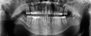
Most authors hypothesize that a visible contour change is caused by a persistent osseous defect of the inferior border of the mandible after BSSO (Figure. 2). These osseous inferior border defects are present in 5.1-36.5% of the sagittal splits one year after BSSO (Agbaje et al., 2013)(Agbaje et al., 2016)(J P Verweij et al., 2017). Known risk factors for these persistent osseous defects of the inferior border include a large mandibular advancement, a large rotation of the occlusal plane, cranial rotation of the proximal segment, and splitting the full thickness of the inferior cortex (Agbaje et al., 2013)(Agbaje et al., 2016)(J P Verweij et al., 2017). However, to our knowledge, no reports are present regarding the association between visible contour changes and osseous inferior border defects. It is unclear in how many percent of cases an osseous defect leads to a visible contour change at the inferior border of the mandible.
The purpose of this study is to determine the incidence of a clinically visible contour change of the inferior mandibular border after BSSO and analyze the association between bony defects and visible contour changes. Possible risk factors for soft tissue contour changes after BSSO are furthermore analyzed.


Study design and inclusion/exclusion criteria
A retrospective cohort study was implemented. The records of consecutive patients who underwent BSSO or a bimaxillary procedure with or without genioplasty to correct a class II malocclusion (mandibular advancement) were reviewed. All patients that received treatment between July 2006 and March 2015 at the department of Oral and Maxillofacial Surgery of the Leiden University Medical Center were included. Patients that received bilateral sagittal split osteotomy to correct a class III malocclusion (mandibular set-back) were not included. Patients were excluded if the follow-up was less than 6 months, if not all radiographs or clinical photographs were present, or if the contour of the mandible could not be assessed by inspection (for example because of a beard).
Variables of interest
The primary outcome of interest in this study was the presence/absence of a visible contour change at the lower border of the mandible after BSSO. A contour change was defined as a deformation of the soft tissue of the inferior border of the mandible, located near the caudal end of the vertical osteotomy site. This was categorized in three groups: (1) no visible contour change; (2) dubious contour change; (3) an evidently visible contour change. Only for group 3, soft tissue contour changes were defined as ‘present’.
Furthermore, a set of specific predictor variables was used to investigate risk factors for visible contour changes at the lower mandibular border. The presence of osseous inferior border defects was defined as a defect in the inferior cortex of the mandible of more than one cortical thickness measured at the osteotomy site. Other predictor variables included presence/absence of third molars during surgery, the occurrence of bad splits, the amount of mandibular movement (mm), the rotation of the occlusal plane (degrees), the postoperative position of the proximal segment, and the pattern of the lingual fracture. The postoperative position of the proximal segment was defined as either a good anatomical position without proximal segment rotation, a slight rotation of the proximal segment (less than one cortex thickness), or significant rotation of the proximal segment (more than one cortex thickness). The lingual fracture pattern was defined as either a type I or type II split. A type I split consisted of a split where the inferior border had split with caudal cortex on both the proximal and distal segment. A type II split consisted of a split that did not run through the caudal cortex, but started in the lingual cortex (including the full thickness of the inferior border), keeping the complete bilateral caudal cortex attached to the proximal segment (J P Verweij et al., 2017).
Data collection
Clinical photographs were taken by a professional medical photographer under standardized conditions before surgery, 6 and 12 months after BSSO. To detect a contour change of the inferior border of the mandible, the postoperative clinical photographs that were taken minimally 6 months after BSSO were analyzed and compared with the preoperative clinical photographs. The defects were diagnosed by one observer and confirmed by two other oral and maxillofacial surgeons.
The occurrence of osseous inferior border defects was analyzed using preoperative radiographs and postoperative radiographs (orthopantomographic images) acquired at the latest follow-up (minimally 6 months after BSSO). A tangential line to the inferior mandibular border was visualized to assess whether a contour change was present near the vertical osteotomy site. This contour change of the inferior mandibular border was measured relative to the thickness of the inferior cortex (i.e., more or less than one cortical thickness) (6).
The presence of third molars and occurrence of bad splits was recorded intra-operatively by the surgeon and noted in the surgical report. The amount of mandibular advancement and rotation of the occlusal plane were calculated after performing cephalometric measurements using the preoperative and postoperative lateral cephalogram. The postoperative proximal segment position was analyzed using the panoramic radiographs or cone-beam computed tomographic images that were performed one week after surgery. The type of split was also analyzed using these images.
The study was performed in accordance with the guidelines of our institution and followed the Declaration of Helsinki on medical protocols and ethics. Because of the retrospective nature of this study, it was granted an exemption by the Leiden University Medical Center institutional review board.
Surgical protocol
BSSO was performed according to the Hunsuck modification (Hunsuck, 1968) using a splitter (Smith Ramus Separator 12 mm, Walter Lorentz Surgical, Jacksonville, FL, USA) and separators (Smith Sagittal Split Separators, curved, Walter Lorentz Surgical, Jacksonville, FL, USA). Six oral and maxillofacial surgeons supervising a resident on the contralateral side used the same surgical technique and surgical protocols, as reported in previous papers (Merkesteyn et al., 2007)(Mensink et al., 2012).
After incision of the mucoperiosteum and exposition of the mandbular bone, the horizontal osteotomy was placed just above the mandibular foramen. The sagittal osteotomy was placed over the anterior side of the ramus ascendence to the distal border of the second molar. The vertical osteotomy was placed just posterior of the second molar, perpendicular to the inferior border of the mandible. An inferior border cut was made completely through the inferior cortex, reaching into the lingual cortex.
The split was performed using splitter and separators. The splitter was placed in the sagittal osteotomy and the separator in the verical osteotomy, in order to guide the split. After a succesful split, the distal segment was placed in the planned position using an intermaxillary wafer. The proximal segment was placed in position using a Luniatschek to secure the condyle in to the mandibular fossa. Simultaneously, the inferior border was palpated to align the inferior border of the distal and proximal segment. Rigid fixation was performed by three bicortical screws.
Follow up included standardized clinical and radiological evaluation and took place at 1 week, 1, 6 and 12 months postoperatively.
Statistical methods
Statistical analyses were performed using the Statistical Package for Social Sciences (SPSS version 23.0 for Mac, IBM, Armonk, NY, USA). Patient characteristics and surgical specifics were reported. To study the association between risk factors and soft tissue defects, generalized linear mixed models (GLMM)(Jiang, 2007) were employed to account for the correlated nature of the data (repeated measurements design consisting of right and left side within one patient). To analyze the association between the presence of a visible soft tissue defect and an osseous inferior border defect the chi-squared test was used.
The medical records of 219 patients, who underwent BSSO-advancement were investigated. Of these patients, 147 could be included in the study. Exclusion was performed because of follow-up of less than six months (10 patients), incomplete radiographs (9 patients) or incomplete or incorrect clinical photographs (53). The mean follow-up time was 398 days (SD 138 days; range 163 - 1164 days). Table 1 shows the patient characteristics.

* Data are represented as number of sites (%).
In the 147 included patients (294 sagittal split sites), the mean mandibular advancement was 5.7mm (range 1-11mm). Third molars were present during the sagittal split in 126 sites (42.9%) and absent in 168 sites (57.1%). Bad splits occured in 8 out of 294 sites (2,7% per site). No bilateral bad splits occurred. Postoperative radiographs showed a significant rotation of the proximal segment in 9 sagittal split sites (3%). Minimal rotation (less than one cortex thickness) was present in 86 sagittal split sites (29.3%). In 199 sides (67,7%) there was no rotatation of the proximal segment. After 257 sagittal splits (87.4%), a type 1 split was present and after 37 sagittal splits (12.6) a type 2 split including the full thickness of the inferior border was present.
An osseous mandibular border defect was present in 21 sagittal split sites (7.1%). Visible contour changes of the soft tissue at the inferior border of the mandible were present in 3 sides (1.0%). An osseous inferior border defect was present in the radiographs of these clinically visible soft tissue contour changers in all 3 patients. In 14,29% of the patients with an osseous mandibular border defect a visible contour change was seen. Table 2 shows the evaluation of soft tissue contour changes.
GLMM were used to study the association between predictor variables and visible contour changes of the inferior mandibular border. There was a statistically significant association between radiographically diagnosed osseous inferior border defects and clinically visible soft tissue contour changes (p lessthan 0.001). No significant associations were recorded for the presence of third molars, the presence of bad splits, the amount of mandibular movement, the rotation of the occlusal plane, the postoperative position of the proximal segment, and the pattern of the lingual fracture with the outcome.

The purpose of this study was to estimate the percentage of a visible contour change of the inferior border of the mandible after BSSO. We furthermore analyzed the association between a clinically visible contour change of the mandible and an osseous inferior border defect and evaluated possible risk factors associated with this clinically visible contour change.
In our study group of 147 patients, a visible contour change after bsso was present in 2% of the patients and 1% of the surgical sides. There was a significant association between the visible contour change of the inferior border of the mandible and an osseous inferior border defect (p lessthan 0.001). No other significant risk factors could be found in this study.
In the current available literature, there is little information available about the incidence of a visible contour change of the inferior border of the mandible after BSSO advancement. Even though many authors have described the contour change as a complication after BSSO, which sometimes can even necessitate secondary reconstruction using bone products or allogeneic implants(Agbaje et al., 2013)(Bouwman et al., 1995)(J P Verweij et al., 2017)(Wolford, 2015). Few studies describe the osseous inferior border defect as a possible cause of a visible contour change, but a correlation was never investigated (Agbaje et al., 2013)(Agbaje et al., 2016)(J P Verweij et al., 2017). In comparison to the osseous inferior border defect, which is present in 5.1-36 percent of the operation sides after BSSO, a visible contour change is quite rare (1-2%).
This is most likely explained by the fact that an osseous inferior border defect is covered by soft tissue that hides the contour changes in most of the cases. Subcutaneous fat can for example fill up the osseous defect. In the older population, the jowl fat pads move caudally towards the inferior border of the mandible and can hide the osseous defect even more (Reece & Rohrich, 2008). In this study group, the vertical osteotomy was placed behind the second molar, and therefore also the masseter muscle could cover the osseous defect, which is typically located near the vertical osteotomy (J P Verweij et al., 2017)(Jop P. Verweij et al., 2015). Possibly an elevated prevalence of visible contour changes of the inferior border of the mandible can be detected after BSSO, when the vertical osteotomy is placed more anteriorly. To enlarge the covering effect of the masseter muscle, Verweij et al. suggested the angled osteotomy concept, where the vertical osteotomy and the inferior border cut is placed more posteriorly (near the masseteric tuberosity). Using this technique, the masseter muscle probably could mask even more of the osseous inferior border defects, but we should notice this study was performed on cadaveric mandibles, so the results must be extrapolated to the clinical setting carefully.
Previous research has identified significant risk factors for osseous inferior border defects in radiographs, including: the amount of advancement, rotation of the occlusal plane, rotation of the proximal segment and whether or not the full thickness of the inferior cortex was completely attached to the proximal mandibular segment (Agbaje et al., 2013)(Agbaje et al., 2016)(J P Verweij et al., 2017). In this study, no significant risk factors (other than the present of an osseous inferior border defect) could be identified for clinically visible inferior border defects or contour changes. This is possibly caused by the low incidence of a visible contour change. Since there is a significant correlation between the osseous defect and a visible contour change, we would expect the same risk factors are valid for a visible contour change, however in this population that could not be proven.
Because of the association between the osseous inferior border defect and a visible contour change, most of the treatment options are aimed at preventing osseous inferior border defects. Wolford et al. and Agbaje et al. described an inferior border osteotomy to perform a split situated in the inferior border to create a continuous inferior border after the advancement (Wolford et al., 1990)(Agbaje et al., 2016). Other authors suggest using bone substitute as grafting material at the osteotomy site in patients treated with large mandibular advancements (>8mm) (Trevisiol et al., 2012)(Raffaini et al., 2020). However, to our knowledge, no reports are present in the current literature that show a lower incidence of visible mandibular border defects after bone grafting during BSSO.
In our clinical experience, visible contour changes are rare after bsso, and even if a contour change is visible, correction is not often necessary. However, when a visible contour change is present after BSSO, the defect can be treated by secondary reconstruction. A possible option is to place bone products or allogeneic implants to fill up the osseous defect and support the soft tissue to improve the visible contour (Agbaje et al., 2013)(J P Verweij et al., 2017)(Wolford, 2015).
We believe our findings represent a reliable estimate of the incidence of soft tissue contour changes in a relatively large group of patients. Some aspects of this retrospective study need to be addressed. The retrospective character of the study and exclusion of 72 patients (because of incomplete data, lack of follow-up or incorrect photographs) is a disadvantage. Several male patients had to be excluded because of wearing a beard and a few female patients had to be excluded because their hair made assessment of the mandible using photographs impossible. There were no remarkable differences between the excluded patient group and our study group, so we therefore do not believe that exclusion influenced any of the results. Clinical inferior border defects were assessed using pre- and postoperative clinical standardized photographs. Although clinical assessment is preferred, we believe this offered a reliable detection method without causing discomfort to patients. All photographs were performed under standardized conditions by a professional medical photographer. Nevertheless, this study simply offers an estimation of the incidence of clinically visible soft tissue contour changes after BSSO. Further research is required to confirm our findings, ideally in a prospective study design including physical examination.
The percentage of a visible contour change of the inferior border of the mandible after BSSO in the present study was found to be 2% per patient and 1% per surgical side. There was a significant association between a visible contour change of the mandible and an osseous inferior border defect (p less than 0.001), however in most cases the osseous defect did not lead to a visible contour change. These findings could help surgeons to estimate the risk developing a visible contour change of the mandible after BSSO.
The authors declare that there are no conflicts of interest in this study.
Clearly Auctoresonline and particularly Psychology and Mental Health Care Journal is dedicated to improving health care services for individuals and populations. The editorial boards' ability to efficiently recognize and share the global importance of health literacy with a variety of stakeholders. Auctoresonline publishing platform can be used to facilitate of optimal client-based services and should be added to health care professionals' repertoire of evidence-based health care resources.

Journal of Clinical Cardiology and Cardiovascular Intervention The submission and review process was adequate. However I think that the publication total value should have been enlightened in early fases. Thank you for all.

Journal of Women Health Care and Issues By the present mail, I want to say thank to you and tour colleagues for facilitating my published article. Specially thank you for the peer review process, support from the editorial office. I appreciate positively the quality of your journal.

Journal of Clinical Research and Reports I would be very delighted to submit my testimonial regarding the reviewer board and the editorial office. The reviewer board were accurate and helpful regarding any modifications for my manuscript. And the editorial office were very helpful and supportive in contacting and monitoring with any update and offering help. It was my pleasure to contribute with your promising Journal and I am looking forward for more collaboration.

We would like to thank the Journal of Thoracic Disease and Cardiothoracic Surgery because of the services they provided us for our articles. The peer-review process was done in a very excellent time manner, and the opinions of the reviewers helped us to improve our manuscript further. The editorial office had an outstanding correspondence with us and guided us in many ways. During a hard time of the pandemic that is affecting every one of us tremendously, the editorial office helped us make everything easier for publishing scientific work. Hope for a more scientific relationship with your Journal.

The peer-review process which consisted high quality queries on the paper. I did answer six reviewers’ questions and comments before the paper was accepted. The support from the editorial office is excellent.

Journal of Neuroscience and Neurological Surgery. I had the experience of publishing a research article recently. The whole process was simple from submission to publication. The reviewers made specific and valuable recommendations and corrections that improved the quality of my publication. I strongly recommend this Journal.

Dr. Katarzyna Byczkowska My testimonial covering: "The peer review process is quick and effective. The support from the editorial office is very professional and friendly. Quality of the Clinical Cardiology and Cardiovascular Interventions is scientific and publishes ground-breaking research on cardiology that is useful for other professionals in the field.

Thank you most sincerely, with regard to the support you have given in relation to the reviewing process and the processing of my article entitled "Large Cell Neuroendocrine Carcinoma of The Prostate Gland: A Review and Update" for publication in your esteemed Journal, Journal of Cancer Research and Cellular Therapeutics". The editorial team has been very supportive.

Testimony of Journal of Clinical Otorhinolaryngology: work with your Reviews has been a educational and constructive experience. The editorial office were very helpful and supportive. It was a pleasure to contribute to your Journal.

Dr. Bernard Terkimbi Utoo, I am happy to publish my scientific work in Journal of Women Health Care and Issues (JWHCI). The manuscript submission was seamless and peer review process was top notch. I was amazed that 4 reviewers worked on the manuscript which made it a highly technical, standard and excellent quality paper. I appreciate the format and consideration for the APC as well as the speed of publication. It is my pleasure to continue with this scientific relationship with the esteem JWHCI.

This is an acknowledgment for peer reviewers, editorial board of Journal of Clinical Research and Reports. They show a lot of consideration for us as publishers for our research article “Evaluation of the different factors associated with side effects of COVID-19 vaccination on medical students, Mutah university, Al-Karak, Jordan”, in a very professional and easy way. This journal is one of outstanding medical journal.

Dear Hao Jiang, to Journal of Nutrition and Food Processing We greatly appreciate the efficient, professional and rapid processing of our paper by your team. If there is anything else we should do, please do not hesitate to let us know. On behalf of my co-authors, we would like to express our great appreciation to editor and reviewers.

As an author who has recently published in the journal "Brain and Neurological Disorders". I am delighted to provide a testimonial on the peer review process, editorial office support, and the overall quality of the journal. The peer review process at Brain and Neurological Disorders is rigorous and meticulous, ensuring that only high-quality, evidence-based research is published. The reviewers are experts in their fields, and their comments and suggestions were constructive and helped improve the quality of my manuscript. The review process was timely and efficient, with clear communication from the editorial office at each stage. The support from the editorial office was exceptional throughout the entire process. The editorial staff was responsive, professional, and always willing to help. They provided valuable guidance on formatting, structure, and ethical considerations, making the submission process seamless. Moreover, they kept me informed about the status of my manuscript and provided timely updates, which made the process less stressful. The journal Brain and Neurological Disorders is of the highest quality, with a strong focus on publishing cutting-edge research in the field of neurology. The articles published in this journal are well-researched, rigorously peer-reviewed, and written by experts in the field. The journal maintains high standards, ensuring that readers are provided with the most up-to-date and reliable information on brain and neurological disorders. In conclusion, I had a wonderful experience publishing in Brain and Neurological Disorders. The peer review process was thorough, the editorial office provided exceptional support, and the journal's quality is second to none. I would highly recommend this journal to any researcher working in the field of neurology and brain disorders.
Dear Agrippa Hilda, Journal of Neuroscience and Neurological Surgery, Editorial Coordinator, I trust this message finds you well. I want to extend my appreciation for considering my article for publication in your esteemed journal. I am pleased to provide a testimonial regarding the peer review process and the support received from your editorial office. The peer review process for my paper was carried out in a highly professional and thorough manner. The feedback and comments provided by the authors were constructive and very useful in improving the quality of the manuscript. This rigorous assessment process undoubtedly contributes to the high standards maintained by your journal.

International Journal of Clinical Case Reports and Reviews. I strongly recommend to consider submitting your work to this high-quality journal. The support and availability of the Editorial staff is outstanding and the review process was both efficient and rigorous.

Thank you very much for publishing my Research Article titled “Comparing Treatment Outcome Of Allergic Rhinitis Patients After Using Fluticasone Nasal Spray And Nasal Douching" in the Journal of Clinical Otorhinolaryngology. As Medical Professionals we are immensely benefited from study of various informative Articles and Papers published in this high quality Journal. I look forward to enriching my knowledge by regular study of the Journal and contribute my future work in the field of ENT through the Journal for use by the medical fraternity. The support from the Editorial office was excellent and very prompt. I also welcome the comments received from the readers of my Research Article.

Dear Erica Kelsey, Editorial Coordinator of Cancer Research and Cellular Therapeutics Our team is very satisfied with the processing of our paper by your journal. That was fast, efficient, rigorous, but without unnecessary complications. We appreciated the very short time between the submission of the paper and its publication on line on your site.

I am very glad to say that the peer review process is very successful and fast and support from the Editorial Office. Therefore, I would like to continue our scientific relationship for a long time. And I especially thank you for your kindly attention towards my article. Have a good day!

"We recently published an article entitled “Influence of beta-Cyclodextrins upon the Degradation of Carbofuran Derivatives under Alkaline Conditions" in the Journal of “Pesticides and Biofertilizers” to show that the cyclodextrins protect the carbamates increasing their half-life time in the presence of basic conditions This will be very helpful to understand carbofuran behaviour in the analytical, agro-environmental and food areas. We greatly appreciated the interaction with the editor and the editorial team; we were particularly well accompanied during the course of the revision process, since all various steps towards publication were short and without delay".

I would like to express my gratitude towards you process of article review and submission. I found this to be very fair and expedient. Your follow up has been excellent. I have many publications in national and international journal and your process has been one of the best so far. Keep up the great work.

We are grateful for this opportunity to provide a glowing recommendation to the Journal of Psychiatry and Psychotherapy. We found that the editorial team were very supportive, helpful, kept us abreast of timelines and over all very professional in nature. The peer review process was rigorous, efficient and constructive that really enhanced our article submission. The experience with this journal remains one of our best ever and we look forward to providing future submissions in the near future.

I am very pleased to serve as EBM of the journal, I hope many years of my experience in stem cells can help the journal from one way or another. As we know, stem cells hold great potential for regenerative medicine, which are mostly used to promote the repair response of diseased, dysfunctional or injured tissue using stem cells or their derivatives. I think Stem Cell Research and Therapeutics International is a great platform to publish and share the understanding towards the biology and translational or clinical application of stem cells.

I would like to give my testimony in the support I have got by the peer review process and to support the editorial office where they were of asset to support young author like me to be encouraged to publish their work in your respected journal and globalize and share knowledge across the globe. I really give my great gratitude to your journal and the peer review including the editorial office.

I am delighted to publish our manuscript entitled "A Perspective on Cocaine Induced Stroke - Its Mechanisms and Management" in the Journal of Neuroscience and Neurological Surgery. The peer review process, support from the editorial office, and quality of the journal are excellent. The manuscripts published are of high quality and of excellent scientific value. I recommend this journal very much to colleagues.

Dr.Tania Muñoz, My experience as researcher and author of a review article in The Journal Clinical Cardiology and Interventions has been very enriching and stimulating. The editorial team is excellent, performs its work with absolute responsibility and delivery. They are proactive, dynamic and receptive to all proposals. Supporting at all times the vast universe of authors who choose them as an option for publication. The team of review specialists, members of the editorial board, are brilliant professionals, with remarkable performance in medical research and scientific methodology. Together they form a frontline team that consolidates the JCCI as a magnificent option for the publication and review of high-level medical articles and broad collective interest. I am honored to be able to share my review article and open to receive all your comments.

“The peer review process of JPMHC is quick and effective. Authors are benefited by good and professional reviewers with huge experience in the field of psychology and mental health. The support from the editorial office is very professional. People to contact to are friendly and happy to help and assist any query authors might have. Quality of the Journal is scientific and publishes ground-breaking research on mental health that is useful for other professionals in the field”.

Dear editorial department: On behalf of our team, I hereby certify the reliability and superiority of the International Journal of Clinical Case Reports and Reviews in the peer review process, editorial support, and journal quality. Firstly, the peer review process of the International Journal of Clinical Case Reports and Reviews is rigorous, fair, transparent, fast, and of high quality. The editorial department invites experts from relevant fields as anonymous reviewers to review all submitted manuscripts. These experts have rich academic backgrounds and experience, and can accurately evaluate the academic quality, originality, and suitability of manuscripts. The editorial department is committed to ensuring the rigor of the peer review process, while also making every effort to ensure a fast review cycle to meet the needs of authors and the academic community. Secondly, the editorial team of the International Journal of Clinical Case Reports and Reviews is composed of a group of senior scholars and professionals with rich experience and professional knowledge in related fields. The editorial department is committed to assisting authors in improving their manuscripts, ensuring their academic accuracy, clarity, and completeness. Editors actively collaborate with authors, providing useful suggestions and feedback to promote the improvement and development of the manuscript. We believe that the support of the editorial department is one of the key factors in ensuring the quality of the journal. Finally, the International Journal of Clinical Case Reports and Reviews is renowned for its high- quality articles and strict academic standards. The editorial department is committed to publishing innovative and academically valuable research results to promote the development and progress of related fields. The International Journal of Clinical Case Reports and Reviews is reasonably priced and ensures excellent service and quality ratio, allowing authors to obtain high-level academic publishing opportunities in an affordable manner. I hereby solemnly declare that the International Journal of Clinical Case Reports and Reviews has a high level of credibility and superiority in terms of peer review process, editorial support, reasonable fees, and journal quality. Sincerely, Rui Tao.

Clinical Cardiology and Cardiovascular Interventions I testity the covering of the peer review process, support from the editorial office, and quality of the journal.

Clinical Cardiology and Cardiovascular Interventions, we deeply appreciate the interest shown in our work and its publication. It has been a true pleasure to collaborate with you. The peer review process, as well as the support provided by the editorial office, have been exceptional, and the quality of the journal is very high, which was a determining factor in our decision to publish with you.

The peer reviewers process is quick and effective, the supports from editorial office is excellent, the quality of journal is high. I would like to collabroate with Internatioanl journal of Clinical Case Reports and Reviews journal clinically in the future time.

Clinical Cardiology and Cardiovascular Interventions, I would like to express my sincerest gratitude for the trust placed in our team for the publication in your journal. It has been a true pleasure to collaborate with you on this project. I am pleased to inform you that both the peer review process and the attention from the editorial coordination have been excellent. Your team has worked with dedication and professionalism to ensure that your publication meets the highest standards of quality. We are confident that this collaboration will result in mutual success, and we are eager to see the fruits of this shared effort.

Dear Dr. Jessica Magne, Editorial Coordinator 0f Clinical Cardiology and Cardiovascular Interventions, I hope this message finds you well. I want to express my utmost gratitude for your excellent work and for the dedication and speed in the publication process of my article titled "Navigating Innovation: Qualitative Insights on Using Technology for Health Education in Acute Coronary Syndrome Patients." I am very satisfied with the peer review process, the support from the editorial office, and the quality of the journal. I hope we can maintain our scientific relationship in the long term.

Dear Monica Gissare, - Editorial Coordinator of Nutrition and Food Processing. ¨My testimony with you is truly professional, with a positive response regarding the follow-up of the article and its review, you took into account my qualities and the importance of the topic¨.

Dear Dr. Jessica Magne, Editorial Coordinator 0f Clinical Cardiology and Cardiovascular Interventions, The review process for the article “The Handling of Anti-aggregants and Anticoagulants in the Oncologic Heart Patient Submitted to Surgery” was extremely rigorous and detailed. From the initial submission to the final acceptance, the editorial team at the “Journal of Clinical Cardiology and Cardiovascular Interventions” demonstrated a high level of professionalism and dedication. The reviewers provided constructive and detailed feedback, which was essential for improving the quality of our work. Communication was always clear and efficient, ensuring that all our questions were promptly addressed. The quality of the “Journal of Clinical Cardiology and Cardiovascular Interventions” is undeniable. It is a peer-reviewed, open-access publication dedicated exclusively to disseminating high-quality research in the field of clinical cardiology and cardiovascular interventions. The journal's impact factor is currently under evaluation, and it is indexed in reputable databases, which further reinforces its credibility and relevance in the scientific field. I highly recommend this journal to researchers looking for a reputable platform to publish their studies.

Dear Editorial Coordinator of the Journal of Nutrition and Food Processing! "I would like to thank the Journal of Nutrition and Food Processing for including and publishing my article. The peer review process was very quick, movement and precise. The Editorial Board has done an extremely conscientious job with much help, valuable comments and advices. I find the journal very valuable from a professional point of view, thank you very much for allowing me to be part of it and I would like to participate in the future!”

Dealing with The Journal of Neurology and Neurological Surgery was very smooth and comprehensive. The office staff took time to address my needs and the response from editors and the office was prompt and fair. I certainly hope to publish with this journal again.Their professionalism is apparent and more than satisfactory. Susan Weiner

My Testimonial Covering as fellowing: Lin-Show Chin. The peer reviewers process is quick and effective, the supports from editorial office is excellent, the quality of journal is high. I would like to collabroate with Internatioanl journal of Clinical Case Reports and Reviews.

My experience publishing in Psychology and Mental Health Care was exceptional. The peer review process was rigorous and constructive, with reviewers providing valuable insights that helped enhance the quality of our work. The editorial team was highly supportive and responsive, making the submission process smooth and efficient. The journal's commitment to high standards and academic rigor makes it a respected platform for quality research. I am grateful for the opportunity to publish in such a reputable journal.

My experience publishing in International Journal of Clinical Case Reports and Reviews was exceptional. I Come forth to Provide a Testimonial Covering the Peer Review Process and the editorial office for the Professional and Impartial Evaluation of the Manuscript.

I would like to offer my testimony in the support. I have received through the peer review process and support the editorial office where they are to support young authors like me, encourage them to publish their work in your esteemed journals, and globalize and share knowledge globally. I really appreciate your journal, peer review, and editorial office.

Dear Agrippa Hilda- Editorial Coordinator of Journal of Neuroscience and Neurological Surgery, "The peer review process was very quick and of high quality, which can also be seen in the articles in the journal. The collaboration with the editorial office was very good."

I would like to express my sincere gratitude for the support and efficiency provided by the editorial office throughout the publication process of my article, “Delayed Vulvar Metastases from Rectal Carcinoma: A Case Report.” I greatly appreciate the assistance and guidance I received from your team, which made the entire process smooth and efficient. The peer review process was thorough and constructive, contributing to the overall quality of the final article. I am very grateful for the high level of professionalism and commitment shown by the editorial staff, and I look forward to maintaining a long-term collaboration with the International Journal of Clinical Case Reports and Reviews.

To Dear Erin Aust, I would like to express my heartfelt appreciation for the opportunity to have my work published in this esteemed journal. The entire publication process was smooth and well-organized, and I am extremely satisfied with the final result. The Editorial Team demonstrated the utmost professionalism, providing prompt and insightful feedback throughout the review process. Their clear communication and constructive suggestions were invaluable in enhancing my manuscript, and their meticulous attention to detail and dedication to quality are truly commendable. Additionally, the support from the Editorial Office was exceptional. From the initial submission to the final publication, I was guided through every step of the process with great care and professionalism. The team's responsiveness and assistance made the entire experience both easy and stress-free. I am also deeply impressed by the quality and reputation of the journal. It is an honor to have my research featured in such a respected publication, and I am confident that it will make a meaningful contribution to the field.

"I am grateful for the opportunity of contributing to [International Journal of Clinical Case Reports and Reviews] and for the rigorous review process that enhances the quality of research published in your esteemed journal. I sincerely appreciate the time and effort of your team who have dedicatedly helped me in improvising changes and modifying my manuscript. The insightful comments and constructive feedback provided have been invaluable in refining and strengthening my work".

I thank the ‘Journal of Clinical Research and Reports’ for accepting this article for publication. This is a rigorously peer reviewed journal which is on all major global scientific data bases. I note the review process was prompt, thorough and professionally critical. It gave us an insight into a number of important scientific/statistical issues. The review prompted us to review the relevant literature again and look at the limitations of the study. The peer reviewers were open, clear in the instructions and the editorial team was very prompt in their communication. This journal certainly publishes quality research articles. I would recommend the journal for any future publications.

Dear Jessica Magne, with gratitude for the joint work. Fast process of receiving and processing the submitted scientific materials in “Clinical Cardiology and Cardiovascular Interventions”. High level of competence of the editors with clear and correct recommendations and ideas for enriching the article.

We found the peer review process quick and positive in its input. The support from the editorial officer has been very agile, always with the intention of improving the article and taking into account our subsequent corrections.

My article, titled 'No Way Out of the Smartphone Epidemic Without Considering the Insights of Brain Research,' has been republished in the International Journal of Clinical Case Reports and Reviews. The review process was seamless and professional, with the editors being both friendly and supportive. I am deeply grateful for their efforts.

To Dear Erin Aust – Editorial Coordinator of Journal of General Medicine and Clinical Practice! I declare that I am absolutely satisfied with your work carried out with great competence in following the manuscript during the various stages from its receipt, during the revision process to the final acceptance for publication. Thank Prof. Elvira Farina

Dear Jessica, and the super professional team of the ‘Clinical Cardiology and Cardiovascular Interventions’ I am sincerely grateful to the coordinated work of the journal team for the no problem with the submission of my manuscript: “Cardiometabolic Disorders in A Pregnant Woman with Severe Preeclampsia on the Background of Morbid Obesity (Case Report).” The review process by 5 experts was fast, and the comments were professional, which made it more specific and academic, and the process of publication and presentation of the article was excellent. I recommend that my colleagues publish articles in this journal, and I am interested in further scientific cooperation. Sincerely and best wishes, Dr. Oleg Golyanovskiy.

Dear Ashley Rosa, Editorial Coordinator of the journal - Psychology and Mental Health Care. " The process of obtaining publication of my article in the Psychology and Mental Health Journal was positive in all areas. The peer review process resulted in a number of valuable comments, the editorial process was collaborative and timely, and the quality of this journal has been quickly noticed, resulting in alternative journals contacting me to publish with them." Warm regards, Susan Anne Smith, PhD. Australian Breastfeeding Association.

Dear Jessica Magne, Editorial Coordinator, Clinical Cardiology and Cardiovascular Interventions, Auctores Publishing LLC. I appreciate the journal (JCCI) editorial office support, the entire team leads were always ready to help, not only on technical front but also on thorough process. Also, I should thank dear reviewers’ attention to detail and creative approach to teach me and bring new insights by their comments. Surely, more discussions and introduction of other hemodynamic devices would provide better prevention and management of shock states. Your efforts and dedication in presenting educational materials in this journal are commendable. Best wishes from, Farahnaz Fallahian.

Dear Maria Emerson, Editorial Coordinator, International Journal of Clinical Case Reports and Reviews, Auctores Publishing LLC. I am delighted to have published our manuscript, "Acute Colonic Pseudo-Obstruction (ACPO): A rare but serious complication following caesarean section." I want to thank the editorial team, especially Maria Emerson, for their prompt review of the manuscript, quick responses to queries, and overall support. Yours sincerely Dr. Victor Olagundoye.

Dear Ashley Rosa, Editorial Coordinator, International Journal of Clinical Case Reports and Reviews. Many thanks for publishing this manuscript after I lost confidence the editors were most helpful, more than other journals Best wishes from, Susan Anne Smith, PhD. Australian Breastfeeding Association.

Dear Agrippa Hilda, Editorial Coordinator, Journal of Neuroscience and Neurological Surgery. The entire process including article submission, review, revision, and publication was extremely easy. The journal editor was prompt and helpful, and the reviewers contributed to the quality of the paper. Thank you so much! Eric Nussbaum, MD

Dr Hala Al Shaikh This is to acknowledge that the peer review process for the article ’ A Novel Gnrh1 Gene Mutation in Four Omani Male Siblings, Presentation and Management ’ sent to the International Journal of Clinical Case Reports and Reviews was quick and smooth. The editorial office was prompt with easy communication.

Dear Erin Aust, Editorial Coordinator, Journal of General Medicine and Clinical Practice. We are pleased to share our experience with the “Journal of General Medicine and Clinical Practice”, following the successful publication of our article. The peer review process was thorough and constructive, helping to improve the clarity and quality of the manuscript. We are especially thankful to Ms. Erin Aust, the Editorial Coordinator, for her prompt communication and continuous support throughout the process. Her professionalism ensured a smooth and efficient publication experience. The journal upholds high editorial standards, and we highly recommend it to fellow researchers seeking a credible platform for their work. Best wishes By, Dr. Rakhi Mishra.

Dear Jessica Magne, Editorial Coordinator, Clinical Cardiology and Cardiovascular Interventions, Auctores Publishing LLC. The peer review process of the journal of Clinical Cardiology and Cardiovascular Interventions was excellent and fast, as was the support of the editorial office and the quality of the journal. Kind regards Walter F. Riesen Prof. Dr. Dr. h.c. Walter F. Riesen.

Dear Ashley Rosa, Editorial Coordinator, International Journal of Clinical Case Reports and Reviews, Auctores Publishing LLC. Thank you for publishing our article, Exploring Clozapine's Efficacy in Managing Aggression: A Multiple Single-Case Study in Forensic Psychiatry in the international journal of clinical case reports and reviews. We found the peer review process very professional and efficient. The comments were constructive, and the whole process was efficient. On behalf of the co-authors, I would like to thank you for publishing this article. With regards, Dr. Jelle R. Lettinga.

Dear Clarissa Eric, Editorial Coordinator, Journal of Clinical Case Reports and Studies, I would like to express my deep admiration for the exceptional professionalism demonstrated by your journal. I am thoroughly impressed by the speed of the editorial process, the substantive and insightful reviews, and the meticulous preparation of the manuscript for publication. Additionally, I greatly appreciate the courteous and immediate responses from your editorial office to all my inquiries. Best Regards, Dariusz Ziora

Dear Chrystine Mejia, Editorial Coordinator, Journal of Neurodegeneration and Neurorehabilitation, Auctores Publishing LLC, We would like to thank the editorial team for the smooth and high-quality communication leading up to the publication of our article in the Journal of Neurodegeneration and Neurorehabilitation. The reviewers have extensive knowledge in the field, and their relevant questions helped to add value to our publication. Kind regards, Dr. Ravi Shrivastava.

Dear Clarissa Eric, Editorial Coordinator, Journal of Clinical Case Reports and Studies, Auctores Publishing LLC, USA Office: +1-(302)-520-2644. I would like to express my sincere appreciation for the efficient and professional handling of my case report by the ‘Journal of Clinical Case Reports and Studies’. The peer review process was not only fast but also highly constructive—the reviewers’ comments were clear, relevant, and greatly helped me improve the quality and clarity of my manuscript. I also received excellent support from the editorial office throughout the process. Communication was smooth and timely, and I felt well guided at every stage, from submission to publication. The overall quality and rigor of the journal are truly commendable. I am pleased to have published my work with Journal of Clinical Case Reports and Studies, and I look forward to future opportunities for collaboration. Sincerely, Aline Tollet, UCLouvain.

Dear Ms. Mayra Duenas, Editorial Coordinator, International Journal of Clinical Case Reports and Reviews. “The International Journal of Clinical Case Reports and Reviews represented the “ideal house” to share with the research community a first experience with the use of the Simeox device for speech rehabilitation. High scientific reputation and attractive website communication were first determinants for the selection of this Journal, and the following submission process exceeded expectations: fast but highly professional peer review, great support by the editorial office, elegant graphic layout. Exactly what a dynamic research team - also composed by allied professionals - needs!" From, Chiara Beccaluva, PT - Italy.

Dear Maria Emerson, Editorial Coordinator, we have deeply appreciated the professionalism demonstrated by the International Journal of Clinical Case Reports and Reviews. The reviewers have extensive knowledge of our field and have been very efficient and fast in supporting the process. I am really looking forward to further collaboration. Thanks. Best regards, Dr. Claudio Ligresti

Dear Chrystine Mejia, Editorial Coordinator, Journal of Neurodegeneration and Neurorehabilitation. “The peer review process was efficient and constructive, and the editorial office provided excellent communication and support throughout. The journal ensures scientific rigor and high editorial standards, while also offering a smooth and timely publication process. We sincerely appreciate the work of the editorial team in facilitating the dissemination of innovative approaches such as the Bonori Method.” Best regards, Dr. Matteo Bonori.
