
Case Report | DOI: https://doi.org/10.31579/2692-9562/050
Associate Professor, Department of ENT, Subbaiah Institute of Medical Sciences, NH-13, Purle, Holebenavalli Post, Shimoga-577222, Karnataka, India.
*Corresponding Author: Sphoorthi Basavannaiah, Associate Professor, Department of ENT, Subbaiah Institute of Medical Sciences, NH-13, Purle, Holebenavalli Post, Shimoga-577222, Karnataka, India.
Citation: Sphoorthi Basavannaiah (2022) Sinister growth behind the ear: how can a person be sloppy and shoddy???Journal of Clinical Otorhinolaryngology 4(2); DOI: 10.31579/2692-9562/050
Copyright: © 2022 Sphoorthi Basavannaiah. This is an open access article distributed under the Creative Commons Attribution License, which permits unrestricted use, distribution, and reproduction in any medium, provided the original work is properly cited.
Received: 15 April 2022 | Accepted: 30 April 2022 | Published: 07 May 2022
Keywords: temporal bone; malignancy; mastoid region; external auditory canal; petrous apex
Malignancy of ear & temporal bone are uncommon and unusual, but if occurs belongs to destructive variety. It can have both local as well as distant spread from external ear canal to inner ear passages. The spread can involve parotid gland to petrous apex. After a prompt radiological investigation, treatment is scheduled as per staging of the disease process. Early detection with treatment will have good prognosis while delayed recognition will have poor prognosis.
Malignancy of ear & temporal bone are rare, aggressive types of tumours. The incidence has been reported to be 1 in 6 cases per million population years, which is < 0>
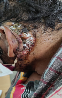
Here, is this adult male aged about 54 years who was encountered in the health camp. He comes with foul smelling discharge from the growth behind the left ear since > 2 months. As per history, the growth was initially size of a pea behind the ear nearly 2 years back which has gradually progressed to have attained the present size. Patient has completely neglected about the growth since the time it has appeared and with no self - hygiene. There is association of pain with this growth occasionally (as per the history from the patient) for which the patient takes analgesics (oral or intramuscular) as and when it pleases him. There was history of otalgia, hearing loss from the left ear. He also gives previous history of recurrent otitis externa taking symptomatic treatment for the same then and there. Patient also gives history of trismus. Patient who is farmer by occupation is a chronic alcoholic, smoker and tobacco/supari /ghutka chewer. There is no h/o any systemic illness.
Patient is normal built and nourished and oriented to time, place and person. His vitals and systemic examination findings are within normal limits. On local ear examination: ulcero-proliferative growth as shown in Figure 1 roughly 4X1 cm seen occupying the left post-auricular region extending vertically from upper edge of pinna upto lower end of lobule of the left ear and horizontally involving the mastoid and temporal bone partly of the left ear. This hard growth has ulcerative margins with areas of necrosis with foul smelling discharge from it. The growth has irregular margins & surface and fixed to the underlying skin. The growth is tender on palpation, sensitive to touch with bleeding from the growth with superficial skin peeling present. The surrounding area of the growth is normal in contour and surface with no induration and with no affect to hair line. Permeatal finding of left ear showed canal erosion and postero-superior retraction of the drum. Biopsy was done under local anaesthesia and later HRCT temporal bone +/- MRI was planned. But the patient did not follow up. The clinical staging in this case was T3. Biopsy report was suggestive of 2 varied diagnosis- Squamous cell carcinoma (as expected most likely) and Haemangioendothelioma (which was just out of the box anticipation).
Tumors of temporal bone include skin cancer of pinna extending to temporal bone, primary tumors of external auditory canal, middle ear, mastoid or petrous apex and metastatic lesions in temporal bone. Primary malignant tumors of temporal bone have an incidence of 0.8-1/10 lakh individuals/year and 60-80% of them are squamous cell carcinoma. Metastatic lesions in temporal bone are rare and originate from breast, lung or kidney tumours [1,3]. Although they can occur at any age, temporal bone tumors are more common in 6th- 7th decade of life and in males. A multifactorial etiology has been suggested for these tumors in whom the risk factors include chronic otitis media, past radiotherapy in the neighbouring regions and exposure to UV radiation (actinic tumours with invasion of EAC) for tumors originating in skin of pinna and EAC, especially in fair-skinned individuals. There can be development of temporal bone carcinoma in patients who have undergone radiotherapy for carcinoma elsewhere in head and neck (i.e nasopharyngeal carcinoma as per Lim et al study). But chronic otitis media has been associated with presence of temporal bone carcinoma with no scientific evidence in etiology till date. Agents such as chlorinated disinfectants or human papillomavirus in cases of carcinomas associated with inverted papillomas have been mentioned as possible carcinogens[2].
Temporal bone tumours manifest with nonspecific symptoms such as otorrhoea, otalgia and/or hearing loss that are often attributed to inflammatory ear diseases. Thus, the ones which have superficial location, diagnosis is often delayed. Tumors of pinna and EAC are known to be more aggressive and have a higher risk of recurrence and lymph node metastasis possibly due to presence of fusion of multiple embryonic planes in this region, which may facilitate tumour dissemination. In addition to clinical examination and histo-pathological analysis, diagnostic imaging assessment of head and neck are essential for accurate tumor diagnosis and staging [4,6]. CT with contrast allows assessing bone erosion and presence of regional adenopathy, whereas MRI with contrast allows a better assessment of its extension to parotid gland, temporomandibular joint, petrous apex and intracranial invasion. In locally advanced tumors, PET allows exclusion of distant metastasis. Currently, there is no universally accepted system for staging of temporal bone carcinoma. The most commonly used is modified Pittsburgh by Moody et al. in 2000 as described below which is based on physical examination, pre-operative CT and presence of facial paralysis [5].
Modified Pittsburgh staging system for temporal bone carcinomas: T1: Tumor limited to EAC without bone erosion, or soft tissue involvement, T2: Tumor with bone erosion limited to EAC (without involving entire thickness) or limited involvement <0>0.5 cm of soft tissues involvement of temporomandibular joint, styloid apophysis or evidence of peripheral facial paralysis [1,6].
Treatment of temporal bone tumours is a challenge for otorhinolaryngologists due to presence of significant neurovascular structures in this region. This usually includes extended tumor surgical resection, which according to its length can be a wide local excision, lateral temporal bone resection, subtotal temporal bone resection or total temporal bone resection. This surgical approach may be combined with cervical dissection with superficial or total parotidectomy and/or supplementary radiotherapy and/or chemotherapy, according to disease extent, presence of lymph node metastases, histological subtype, available resources, and surgeon’s inclination [2,3,4,5].
Temporal bone malignancies are rare often presenting in the setting of long standing chronic otitis media and often at an advanced stage. The tissue diagnosis is relatively forthright but can be tricky sometimes. However, staging the disease is a intricate task that is best approached with consideration of triad features that is clinical, radiological and pathological findings. The evidence based management of these uncommon tumors is not well established and as such standardisation of surgical and adjuvant treatment, as well as pathological reporting will contribute to more clear management pathways in the future.
It was my pleasure to submit my testimonial concerning the Reviewer Board of our Scientific Journal “Brain and Neurological Disorders”. The Reviewers focused on some modifications and their contribution was helpful. The ladies of our Editorial Office were also supported my efforts. It was my honor to have such a co-operation and I am looking forward for more collaboration.

Dear Grace Pierce, Editorial Coordinator of Journal of Clinical Research and Reports, Thank you for the speedy and efficient peer review process. I appreciate the fact that your peer reviewers do not take months to respond like with some other journals. I would also like to thank the editorial office for responding quickly to my questions. It is an excellent journal. I plan to submit more manuscripts in the future. Best wishes from, Robert W. McGee

Dear Grace Pierce, Editorial Coordinator of Journal of Clinical Research and Reports, Working with you and your team on our recent publication in JCRR has been a truly wonderful and enjoyable experience. The responses were prompt, and the reviewers were patient, constructive, and highly professional. One reviewer in particular gave me the feeling that a professor was carefully reading and commenting on my coursework, which was deeply touching. The entire process was straightforward and hassle‑free, with no tedious online forms to complete. I highly recommend this journal. Best wishes from, DR Aibing Rao, Head of R&D

I Appreciate the Opportunity to Share my Experience with the Journal of Clinical Research and Reports. The peer review process was timely and constructive, and the feedback provided helped improve the quality of our manuscript. The editorial office was professional, responsive, and supportive throughout the process, ensuring smooth communication and efficient handling of the submission. Overall, it was a positive experience collaborating with your team.

Dear Mercy Grace, Editorial Coordinator of Obstetrics Gynecology and Reproductive Sciences, We would like to express our gratitude for your help at all stages of publishing and editing the article. The editors of the magazine answer all the necessary questions and help at every stage. We will definitely continue to cooperate and publish other works in the Obstetrics Gynecology and Reproductive Sciences! Best wishes from, Alla Konstantinovna Politova,

Dear Maria Emerson, Editorial Coordinator of International Journal of Clinical Case Reports and Reviews, What distinguishes International Journal of Clinical Case Report and Review is not only the scientific rigor of its publications, but the intellectual climate in which research is evaluated. The submission process is refreshingly free of unnecessary formal barriers and bureaucratic rituals that often complicate academic publishing without adding real value. The peer-review system is demanding yet constructive, guided by genuine scientific dialogue rather than hierarchical or authoritarian attitudes. Reviewers act as collaborators in improving the manuscript, not as gatekeepers imposing arbitrary standards. This journal offers a rare balance: high methodological standards combined with a respectful, transparent, and supportive editorial approach. In an era where publishing can feel more burdensome than research itself, this platform restores the original purpose of peer review — to refine ideas, not to obstruct them Prof. Perlat Kapisyzi, FCCP PULMONOLOGIST AND THORACIC IMAGING.
