AUCTORES
Globalize your Research

Research Article | DOI: https://doi.org/10.31579/2642-9756/017
*Corresponding Author: Nagy M. Metwally Ahmed, Lecturer of Obstetrics and Gynecology, Obstetrics and Gynecology Department, Faculty of Medicine; Zagazig University. Zagazig, Egypt.
Citation: Metwally Ahmed NM. (2020) Evaluation of risk of premalignant and malignant changes of the cervix in systemic lupus erythromatosis patientss. J Women Health Care Issues, 3(1); Doi/10.31579/2642-9756/017
Copyright: ©2020. Nagy M. Metwally Ahmed, This is an open-access article distributed under the terms of the Creative Commons Attribution License, which permits unrestricted use, distribution, and reproduction in any medium, provided the original author and source are credited.
Received: 16 December 2019 | Accepted: 02 January 2020 | Published: 07 January 2020
Keywords: premalignancy; malignant changes; cervix
Introduction: Cervical cancer is the second most common malignancy among women all over the world and a leading cause of death in females. There are known risk factors in this regard including immunosuppression condition like SLE. Screening of women could decrease the incidence of cervical cancer dramatically by detection of early premalignant changes in the tissue. There was significant debate as female with SLE did in fact have an increased risk of cancer compared to the general population. Our study was conducted to determine the prevalence of abnormal cervical smear and colposcopy finding in patients with SLE.
Patients and methods: Our study was carried out on 128 participants (64 SLE patients and 64 apparently healthy individuals as a control group). Patients were recruited from Rheumatology, Obstetrics and Gynecology Department of Armed Forces Hospitals of South Region. Gazan, KSA. The study was explained to all patients and informed consent was obtained before enrollment in the study. All patients underwent thorough complete history taking, clinical evaluation, local and bimanual examination, speculum examination with pap smear intake, colposcopy examination to whom have abnormal pap smear patients with suspicious area, multiple biopsies where be taken for histopathologic examination.
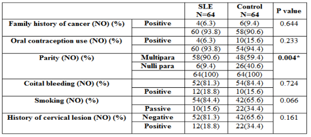
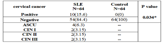
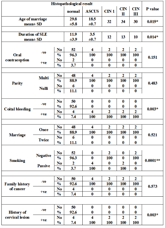
Results: The results of our study demonstrated that, there is a significant association between cervical cancer and SLE patients (p=0.034). There is a significant association between ASCUS (Atypical squamous cells of undetermined significance) with younger age of marriage (p=0.019) and shorter duration of SLE (0.014). There is a significant association between cervical cancer with coital bleeding and history of cervical lesion (p=0.003). There is a high significant association between cervical cancer (especially ASCUS and CIN II) with passive smoking. Other examined parameters did not show any significant association (p>0.05).
Although the overall mortality of patients with carcinoma of the cervix has decreased over the years on account of the widespread availability of effective screening programs, cervical cancer is the second most common malignancy in women and a major cause of morbidity and mortality, It an approximately represented by 510,000 newly diagnosed cases and 288,000 related deaths. Overall, the 5-year survival rate has been reported to be 73%, but the prognosis unsatisfactory [1].
Cervical cancer was recognized for decades to behave as a sexually transmitted disease, Sexually transmitted HPV infection was implicated in its pathogenesis. In the mid-1970s, Meisels and Fortin recognized, on morphologic grounds, that HPV infection of the cervix occurred frequently, often with the histological characteristics of mild CIN [2].
Systemic lupus erythematosus (SLE), or lupus, is a chronic, progressive, autoimmune disorder that affects multiple organ systems, with a broad range of clinical and laboratory manifestations. The term lupus means wolf in Latin and is named as such because of facial lesions found in the disease process that are reminiscent of a wolf ’s bite [3].
An important complication of SLE is increased incidence of malignancy including cervical cancers, autoimmune system dysfunction, recurrent infections.The immunosuppressive medication exposure may constitute explanations which suggest increased incidence of malignancy in SLE [4].
Cytology has been applied to the diagnosis of HPV infections of the penis, vulva, anus, and cervix through (Pap smear). The Papanicolaou-stained (Pap) smear is the most common method for detecting high-risk HPV. This method was named after pathologist George Papanicolaou, who pioneered this method in 1949, before HPV was implicated in cervical cancer. It is a screening tool for morphological changes in cervical cells [5].
Determined the occurrence of cervical dysplasia and Pap smear abnormalities in patients with systemic lupus erythematosus. The demonstrated that patients with SLE have more risk of cervical dysplasia than general population; they suggested that epidemiological surveillance of this group of patients should be more careful, with pap smear more early in life and probably every 6 months apart [6].
Studied the prevalence of abnormal cervicovaginal smears in patients with systemic lupus erythematosus. They concluded that cervicovaginal smearing is an easy, economic, safe, repeatable and noninvasive technique for screening and early detection of cervical neoplastic lesions in SLE [7].
The aim of this study is to determine the relationship between systemic lupus erythematosus and premalignant and malignant changes of cervix
Subjects and methods:
The subjects;
A cross sectional case control study was conducted on 128 include 64 SLE patients. The ages of collected cases ranged from 22 to 43 years (mean±SD31.5±6.2years). The disease duration ranged from 1.5 to 16 years (mean±SD11.1±4.9 years). In addition to this study include 64 healthy volunteers not known to suffer from any rheumatological manifestations as control group. All cases were collected during the period (from June 2017 to February 2019)
These patients were collected from Rheumatology, gynecology and obstetrics department, Armed Forces Hospitals of South Region. Gazan KSA
Inclusion criteria:
Exclusion criteria:
Design of the study;
All patients were subjected to the followings:
Both patients and control group were subjected to the following
Written informed consents were obtained from each patient participating in this study after informing them about the steps of study.
Each participant was subjected according to guidelines in 2013:
Statistical analysis
Data entry and statistical analyses were performed using SPSS (statistical package of social sciences) version 21 (SPSS Inc., Chicago, IL, Categorical data were expressed in number and percentage. Continuous normally distributed data were expressed in mean and standard deviation (10). The quantitative data were examined by Kolmogrov Smirnov test for normality of data [11]. Statistical significance was considered when probability (P) value was less than or equal to 0.05.
Student T test was used for continuous normally distributed data. To study the association between categorical data in SLE patients with those of the control group using chi square test or fisher exact test. Comparison of continuous data with various types of cancer cervix was conducted ANOVA (analysis of variance).
A case control study was conducted on 64 SLE female patients as well as 64 females' healthy volunteers were selected as control group. There was no statistically significant difference between SLE and control group regarding the Demographic characteristics. The results of our study demonstrated that, there is a significant association between cervical cancer and SLE patients (p=0.034). There is a significant association between ASCUS with younger age of marriage (p=0.019) and shorter duration of SLE (0.014) .
There is a significant association between cervical cancer with coital bleeding and history of cervical lesion (p=0.003). There is a high significant association between cervical cancer (especially ASCUS and CIN II) with passive smoking. Other examined parameters did not show any significant association (p>0.05).
In our study, we investigated the Performance values of Pap smear and colposcopy in comparison with histopathology results, pap smear had 60%sensitivity, 100%specificity, 100% positive predictive value, 93.1% negative predictive value. While colposcopy showed higher results 80% sensitivity, 100%specificity, 100% positive predictive value, 96.4% negative predictive value.

*p<0.05 is significant
**p<0.0001 is highly significant
ASCUS atypical squamous cervical undetermined significance
CIN I cervical interepithelial lesion I
CIN II cervical interepithelial lesion II
CIN III cervical interepithelial lesion III
Table 4: Association between premalignant changes of cervix and clinical characteristics of SLE patients

Table 5: Association between histopathology, pap smear and colposcopy
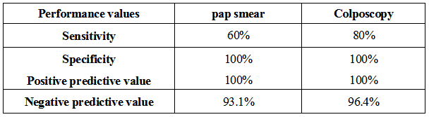
Table 6: Performance values of Pap smear and colposcopy in comparison with histopathology results
Cervical cancer is the second most commonly occurring cancer in women and accounts for up to 300 000 annual deaths [12]. Overall, the 5-year survival rate has been reported to be 73%, but the prognosis unsatisfactory [13].
The causes of cervical cancer in various sources includes early sexual activity, multiple sexual partners, HPV infection, genital warts, sexually transmitted diseases, genital tract abnormalities, age, smoking, passive smoke, poor nutrition, immunodeficiency and malnutrition. [14].
Several studies have shown a possible link between SLE and cervical cancer. Along with a high infection rate of HPV in SLE patients, many studies have shown an increased incidence of cervical cancer in SLE patients. Moreover, other studies have noted a high risk of abnormal Pap tests in women with SLE [15].
SLE is a multi-organ system, autoimmune disease with numerous immunological and clinical manifestations. It is believed to develop as a result of dysregulation of the immune system, ultimately leading to the clinical features of inflammation [15]. Also, it was reported an association between immunological disorder and cervical premalignant and malignant abnormalities [16, 17].
In addition to the clinical manifestations of SLE itself, female patients also have to contend with a heightened risk of developing abnormal cervical smears and squamous intra-epithelial lesions of the cervix as well as other cancers [18].
Interesting researches have shown that SLE syndrome represents a permissive effect of immunosuppression on increased host susceptibility to high cancer-risk and infections, the causative agents of CIN and cervical cancer [19].
Our study was conducted to determine the prevalence of abnormal cervical smear and colposcopic finding in patients with SLE. Ourcase control study was designed on64 SLE female patients as well as 64 females healthy volunteers were selected as control group.
In this study, there is a significant association between cervical cancer and SLE patients (p=0.034). 4 patients (6.3%) of SLE had ASCUS while 2patients (3.1%) of SLE had CIN I type as well as 2 patients (3.1%) of SLE had CIN II, 2 patients (3.1%) of SLE had CIN III .
We confirm recent results by Cao and his colleagues in 2015 who detected high significant association of cervical cancer and SLE patients and reported that cervical cancer was one of common cancers between SLE patients [18].
We also in harmony with earlier worldwide results of They had demonstrated cervical neoplasia among common cancers on top of SLE disease [20].
Moreover, we agree with a large meta-analysis research by Liu and hiscoworkers applied on 2011, there was a large difference between case and control groups with an increased risk of cervical neoplasia with SLE. The consensus in the literature suggests that immunosuppressive treatment probably does contribute to the development of cervical disease in patients with SLE as well as the effect of the disease itself [21].
Interestingly, we share recent Egyptian research results of Al- who detected and studied the prevalence of abnormal cervical smears in SLE patients, they conducted that cervical smear is easy, safe and noninvasive technique for screening and early detection of cervical neoplasia in SLE patients. Among these 4 positive patients, 3 were having ASCUS and one was having LSIL while negative results were detected in control group [7].
We also agree with previous study, they reported that SLE itself was identified as independent risk factors for high risk HPV infection among Korean women along with ≥2 sexual partners. High-risk HPV infection and cervical cytological abnormalities were more common among Korean women with SLE than controls. They concluded that SLE itself can be a risk factor for HPV infection among Korean women, suggesting the importance of close monitoring of cervical cytology in SLE [22].
Also, they detected epithelial dysplasia in 8 patients, atypical squamous cells of undetermined significance (ASCUS) in 7 patients, and carcinoma in situ (CIN) in 7 patients.
We also agree with the most recent study in April 2017 by they Based on 121 events of cervical neoplasia during 23 136 person-years among SLE patients, there was an increased risk of any cervical neoplasia compared with the general population, but not invasive cervical cancer [23].
The subcohort treated with other immunosuppressants was at highest risk of cervical neoplasia.
Although we could not evaluate the immunosuppressive treatment role in cervical cancer incidence due to short time of study and lake of follow up period, however we found a disagreement among different studies regarding the role of immunosuppressive drugs as a predisposing factor for these cervical abnormalities. On one side, most of the studies showed no association between the use of immunosuppressive agents and increased frequency of cervical abnormalities, while two others have suggested the participation of intravenous cyclophosphamideon the development of this cervical complication. [24]
In our study, when we studied clinical characteristics and type of cervical cancer, there is a significant association between ASCUS with younger age of marriage (p=0.019) and shorter duration of SLE (p=0.014). While, other studied parameters did not show any significant association.
We are in disharmony with a study could not detect any significant association between duration of SLE and cancer development although they had long period of follow up [25].
We did not detect a significant association between any type of cervical cancer and oral contraception use in SLE patients. Which disagree with who reported oral contraceptive pills as a possible risk factor in cervical cancer. This conflict can be due to different in number of study participants and higher number of cervical cancers SLE patients in the later study [26].
In this study, there is a high significant association between cervical cancer (especially ASCUS and CIN II) with passive smoking. This result was not coenside with earlier research on malignancy on top of SLE; who highlighted smoking as high-risk factor in lung cancer rather than cervical cancer [27].
We also in disagreement with who detected smoking in a similar ratio between SLE patients and control group with no significant difference [28].
Previous studies have demonstrated an increased prevalence of atypical cervical smears in patients with SLE even in asymptomatic patients, ranging from 24 to 36%, compared with a prevalence of ≤5–15% in controls [29].
Pap smear abnormalities in female patients with autoimmune diseases had been previously investigated. Pap smear screening was done for autoimmune group include SLE, Rheumatoid arthritis, ankylosing arthritis and systemic sclerosis compared with normal counterparts [17].
The two groups were matched for demographics and known risk factors of cervical malignancy. Frequency of abnormal Pap smear testing was significantly higher in the case group comparing with that in the controls. Frequency of abnormal Pap smear testing was higher in the patients with SLE and RA comparing with that in the controls [17].
In our study, we investigated the Performance values of Pap smear and colposcopy in comparison with histopathology results, pap smear had 60%sensitivity, 100%specificity, 100% positive predictive value, 93.1% negative predictive value. While colposcopy showed higher results 80% sensitivity, 100%specificity, 100% positive predictive value, 96.4% negative predictive value.
This come with accordance with the result of who detected sensitivity in 60%of pap smear, 87%specificity, 43% positive predictive value, 93% negative predictive value with higher values regarding colposcopy. It had 75%sensitivity, 99%specificity, 94% positive predictive value, 96% negative predictive value (30).
Determining accurate cancer risk in a disease like SLE, which nowadays may affect patients for several decades, is very challenging. While, mortality and morbidity were higher in SLE patients as well as high incidence of cancer elevated with long standing SLE, regular screening investigation must be applied for all SLE patients especially who had possible risk factors. Colposcopy show higher specificity and sensitivity
Recommendations
Patients with SLE had higher risk for cervical dysplasia than general population. Thus, gynecological visits at shorter intervals (3-6months) recommended for these patients especially those with previous history of younger age of marriage, cervical lesion, with coital bleeding and with passive smoking.
Limitation:
Clearly Auctoresonline and particularly Psychology and Mental Health Care Journal is dedicated to improving health care services for individuals and populations. The editorial boards' ability to efficiently recognize and share the global importance of health literacy with a variety of stakeholders. Auctoresonline publishing platform can be used to facilitate of optimal client-based services and should be added to health care professionals' repertoire of evidence-based health care resources.

Journal of Clinical Cardiology and Cardiovascular Intervention The submission and review process was adequate. However I think that the publication total value should have been enlightened in early fases. Thank you for all.

Journal of Women Health Care and Issues By the present mail, I want to say thank to you and tour colleagues for facilitating my published article. Specially thank you for the peer review process, support from the editorial office. I appreciate positively the quality of your journal.

Journal of Clinical Research and Reports I would be very delighted to submit my testimonial regarding the reviewer board and the editorial office. The reviewer board were accurate and helpful regarding any modifications for my manuscript. And the editorial office were very helpful and supportive in contacting and monitoring with any update and offering help. It was my pleasure to contribute with your promising Journal and I am looking forward for more collaboration.

We would like to thank the Journal of Thoracic Disease and Cardiothoracic Surgery because of the services they provided us for our articles. The peer-review process was done in a very excellent time manner, and the opinions of the reviewers helped us to improve our manuscript further. The editorial office had an outstanding correspondence with us and guided us in many ways. During a hard time of the pandemic that is affecting every one of us tremendously, the editorial office helped us make everything easier for publishing scientific work. Hope for a more scientific relationship with your Journal.

The peer-review process which consisted high quality queries on the paper. I did answer six reviewers’ questions and comments before the paper was accepted. The support from the editorial office is excellent.

Journal of Neuroscience and Neurological Surgery. I had the experience of publishing a research article recently. The whole process was simple from submission to publication. The reviewers made specific and valuable recommendations and corrections that improved the quality of my publication. I strongly recommend this Journal.

Dr. Katarzyna Byczkowska My testimonial covering: "The peer review process is quick and effective. The support from the editorial office is very professional and friendly. Quality of the Clinical Cardiology and Cardiovascular Interventions is scientific and publishes ground-breaking research on cardiology that is useful for other professionals in the field.

Thank you most sincerely, with regard to the support you have given in relation to the reviewing process and the processing of my article entitled "Large Cell Neuroendocrine Carcinoma of The Prostate Gland: A Review and Update" for publication in your esteemed Journal, Journal of Cancer Research and Cellular Therapeutics". The editorial team has been very supportive.

Testimony of Journal of Clinical Otorhinolaryngology: work with your Reviews has been a educational and constructive experience. The editorial office were very helpful and supportive. It was a pleasure to contribute to your Journal.

Dr. Bernard Terkimbi Utoo, I am happy to publish my scientific work in Journal of Women Health Care and Issues (JWHCI). The manuscript submission was seamless and peer review process was top notch. I was amazed that 4 reviewers worked on the manuscript which made it a highly technical, standard and excellent quality paper. I appreciate the format and consideration for the APC as well as the speed of publication. It is my pleasure to continue with this scientific relationship with the esteem JWHCI.

This is an acknowledgment for peer reviewers, editorial board of Journal of Clinical Research and Reports. They show a lot of consideration for us as publishers for our research article “Evaluation of the different factors associated with side effects of COVID-19 vaccination on medical students, Mutah university, Al-Karak, Jordan”, in a very professional and easy way. This journal is one of outstanding medical journal.

Dear Hao Jiang, to Journal of Nutrition and Food Processing We greatly appreciate the efficient, professional and rapid processing of our paper by your team. If there is anything else we should do, please do not hesitate to let us know. On behalf of my co-authors, we would like to express our great appreciation to editor and reviewers.

As an author who has recently published in the journal "Brain and Neurological Disorders". I am delighted to provide a testimonial on the peer review process, editorial office support, and the overall quality of the journal. The peer review process at Brain and Neurological Disorders is rigorous and meticulous, ensuring that only high-quality, evidence-based research is published. The reviewers are experts in their fields, and their comments and suggestions were constructive and helped improve the quality of my manuscript. The review process was timely and efficient, with clear communication from the editorial office at each stage. The support from the editorial office was exceptional throughout the entire process. The editorial staff was responsive, professional, and always willing to help. They provided valuable guidance on formatting, structure, and ethical considerations, making the submission process seamless. Moreover, they kept me informed about the status of my manuscript and provided timely updates, which made the process less stressful. The journal Brain and Neurological Disorders is of the highest quality, with a strong focus on publishing cutting-edge research in the field of neurology. The articles published in this journal are well-researched, rigorously peer-reviewed, and written by experts in the field. The journal maintains high standards, ensuring that readers are provided with the most up-to-date and reliable information on brain and neurological disorders. In conclusion, I had a wonderful experience publishing in Brain and Neurological Disorders. The peer review process was thorough, the editorial office provided exceptional support, and the journal's quality is second to none. I would highly recommend this journal to any researcher working in the field of neurology and brain disorders.
Dear Agrippa Hilda, Journal of Neuroscience and Neurological Surgery, Editorial Coordinator, I trust this message finds you well. I want to extend my appreciation for considering my article for publication in your esteemed journal. I am pleased to provide a testimonial regarding the peer review process and the support received from your editorial office. The peer review process for my paper was carried out in a highly professional and thorough manner. The feedback and comments provided by the authors were constructive and very useful in improving the quality of the manuscript. This rigorous assessment process undoubtedly contributes to the high standards maintained by your journal.

International Journal of Clinical Case Reports and Reviews. I strongly recommend to consider submitting your work to this high-quality journal. The support and availability of the Editorial staff is outstanding and the review process was both efficient and rigorous.

Thank you very much for publishing my Research Article titled “Comparing Treatment Outcome Of Allergic Rhinitis Patients After Using Fluticasone Nasal Spray And Nasal Douching" in the Journal of Clinical Otorhinolaryngology. As Medical Professionals we are immensely benefited from study of various informative Articles and Papers published in this high quality Journal. I look forward to enriching my knowledge by regular study of the Journal and contribute my future work in the field of ENT through the Journal for use by the medical fraternity. The support from the Editorial office was excellent and very prompt. I also welcome the comments received from the readers of my Research Article.

Dear Erica Kelsey, Editorial Coordinator of Cancer Research and Cellular Therapeutics Our team is very satisfied with the processing of our paper by your journal. That was fast, efficient, rigorous, but without unnecessary complications. We appreciated the very short time between the submission of the paper and its publication on line on your site.

I am very glad to say that the peer review process is very successful and fast and support from the Editorial Office. Therefore, I would like to continue our scientific relationship for a long time. And I especially thank you for your kindly attention towards my article. Have a good day!

"We recently published an article entitled “Influence of beta-Cyclodextrins upon the Degradation of Carbofuran Derivatives under Alkaline Conditions" in the Journal of “Pesticides and Biofertilizers” to show that the cyclodextrins protect the carbamates increasing their half-life time in the presence of basic conditions This will be very helpful to understand carbofuran behaviour in the analytical, agro-environmental and food areas. We greatly appreciated the interaction with the editor and the editorial team; we were particularly well accompanied during the course of the revision process, since all various steps towards publication were short and without delay".

I would like to express my gratitude towards you process of article review and submission. I found this to be very fair and expedient. Your follow up has been excellent. I have many publications in national and international journal and your process has been one of the best so far. Keep up the great work.

We are grateful for this opportunity to provide a glowing recommendation to the Journal of Psychiatry and Psychotherapy. We found that the editorial team were very supportive, helpful, kept us abreast of timelines and over all very professional in nature. The peer review process was rigorous, efficient and constructive that really enhanced our article submission. The experience with this journal remains one of our best ever and we look forward to providing future submissions in the near future.

I am very pleased to serve as EBM of the journal, I hope many years of my experience in stem cells can help the journal from one way or another. As we know, stem cells hold great potential for regenerative medicine, which are mostly used to promote the repair response of diseased, dysfunctional or injured tissue using stem cells or their derivatives. I think Stem Cell Research and Therapeutics International is a great platform to publish and share the understanding towards the biology and translational or clinical application of stem cells.

I would like to give my testimony in the support I have got by the peer review process and to support the editorial office where they were of asset to support young author like me to be encouraged to publish their work in your respected journal and globalize and share knowledge across the globe. I really give my great gratitude to your journal and the peer review including the editorial office.

I am delighted to publish our manuscript entitled "A Perspective on Cocaine Induced Stroke - Its Mechanisms and Management" in the Journal of Neuroscience and Neurological Surgery. The peer review process, support from the editorial office, and quality of the journal are excellent. The manuscripts published are of high quality and of excellent scientific value. I recommend this journal very much to colleagues.

Dr.Tania Muñoz, My experience as researcher and author of a review article in The Journal Clinical Cardiology and Interventions has been very enriching and stimulating. The editorial team is excellent, performs its work with absolute responsibility and delivery. They are proactive, dynamic and receptive to all proposals. Supporting at all times the vast universe of authors who choose them as an option for publication. The team of review specialists, members of the editorial board, are brilliant professionals, with remarkable performance in medical research and scientific methodology. Together they form a frontline team that consolidates the JCCI as a magnificent option for the publication and review of high-level medical articles and broad collective interest. I am honored to be able to share my review article and open to receive all your comments.

“The peer review process of JPMHC is quick and effective. Authors are benefited by good and professional reviewers with huge experience in the field of psychology and mental health. The support from the editorial office is very professional. People to contact to are friendly and happy to help and assist any query authors might have. Quality of the Journal is scientific and publishes ground-breaking research on mental health that is useful for other professionals in the field”.

Dear editorial department: On behalf of our team, I hereby certify the reliability and superiority of the International Journal of Clinical Case Reports and Reviews in the peer review process, editorial support, and journal quality. Firstly, the peer review process of the International Journal of Clinical Case Reports and Reviews is rigorous, fair, transparent, fast, and of high quality. The editorial department invites experts from relevant fields as anonymous reviewers to review all submitted manuscripts. These experts have rich academic backgrounds and experience, and can accurately evaluate the academic quality, originality, and suitability of manuscripts. The editorial department is committed to ensuring the rigor of the peer review process, while also making every effort to ensure a fast review cycle to meet the needs of authors and the academic community. Secondly, the editorial team of the International Journal of Clinical Case Reports and Reviews is composed of a group of senior scholars and professionals with rich experience and professional knowledge in related fields. The editorial department is committed to assisting authors in improving their manuscripts, ensuring their academic accuracy, clarity, and completeness. Editors actively collaborate with authors, providing useful suggestions and feedback to promote the improvement and development of the manuscript. We believe that the support of the editorial department is one of the key factors in ensuring the quality of the journal. Finally, the International Journal of Clinical Case Reports and Reviews is renowned for its high- quality articles and strict academic standards. The editorial department is committed to publishing innovative and academically valuable research results to promote the development and progress of related fields. The International Journal of Clinical Case Reports and Reviews is reasonably priced and ensures excellent service and quality ratio, allowing authors to obtain high-level academic publishing opportunities in an affordable manner. I hereby solemnly declare that the International Journal of Clinical Case Reports and Reviews has a high level of credibility and superiority in terms of peer review process, editorial support, reasonable fees, and journal quality. Sincerely, Rui Tao.

Clinical Cardiology and Cardiovascular Interventions I testity the covering of the peer review process, support from the editorial office, and quality of the journal.

Clinical Cardiology and Cardiovascular Interventions, we deeply appreciate the interest shown in our work and its publication. It has been a true pleasure to collaborate with you. The peer review process, as well as the support provided by the editorial office, have been exceptional, and the quality of the journal is very high, which was a determining factor in our decision to publish with you.

The peer reviewers process is quick and effective, the supports from editorial office is excellent, the quality of journal is high. I would like to collabroate with Internatioanl journal of Clinical Case Reports and Reviews journal clinically in the future time.

Clinical Cardiology and Cardiovascular Interventions, I would like to express my sincerest gratitude for the trust placed in our team for the publication in your journal. It has been a true pleasure to collaborate with you on this project. I am pleased to inform you that both the peer review process and the attention from the editorial coordination have been excellent. Your team has worked with dedication and professionalism to ensure that your publication meets the highest standards of quality. We are confident that this collaboration will result in mutual success, and we are eager to see the fruits of this shared effort.

Dear Dr. Jessica Magne, Editorial Coordinator 0f Clinical Cardiology and Cardiovascular Interventions, I hope this message finds you well. I want to express my utmost gratitude for your excellent work and for the dedication and speed in the publication process of my article titled "Navigating Innovation: Qualitative Insights on Using Technology for Health Education in Acute Coronary Syndrome Patients." I am very satisfied with the peer review process, the support from the editorial office, and the quality of the journal. I hope we can maintain our scientific relationship in the long term.

Dear Monica Gissare, - Editorial Coordinator of Nutrition and Food Processing. ¨My testimony with you is truly professional, with a positive response regarding the follow-up of the article and its review, you took into account my qualities and the importance of the topic¨.

Dear Dr. Jessica Magne, Editorial Coordinator 0f Clinical Cardiology and Cardiovascular Interventions, The review process for the article “The Handling of Anti-aggregants and Anticoagulants in the Oncologic Heart Patient Submitted to Surgery” was extremely rigorous and detailed. From the initial submission to the final acceptance, the editorial team at the “Journal of Clinical Cardiology and Cardiovascular Interventions” demonstrated a high level of professionalism and dedication. The reviewers provided constructive and detailed feedback, which was essential for improving the quality of our work. Communication was always clear and efficient, ensuring that all our questions were promptly addressed. The quality of the “Journal of Clinical Cardiology and Cardiovascular Interventions” is undeniable. It is a peer-reviewed, open-access publication dedicated exclusively to disseminating high-quality research in the field of clinical cardiology and cardiovascular interventions. The journal's impact factor is currently under evaluation, and it is indexed in reputable databases, which further reinforces its credibility and relevance in the scientific field. I highly recommend this journal to researchers looking for a reputable platform to publish their studies.

Dear Editorial Coordinator of the Journal of Nutrition and Food Processing! "I would like to thank the Journal of Nutrition and Food Processing for including and publishing my article. The peer review process was very quick, movement and precise. The Editorial Board has done an extremely conscientious job with much help, valuable comments and advices. I find the journal very valuable from a professional point of view, thank you very much for allowing me to be part of it and I would like to participate in the future!”

Dealing with The Journal of Neurology and Neurological Surgery was very smooth and comprehensive. The office staff took time to address my needs and the response from editors and the office was prompt and fair. I certainly hope to publish with this journal again.Their professionalism is apparent and more than satisfactory. Susan Weiner

My Testimonial Covering as fellowing: Lin-Show Chin. The peer reviewers process is quick and effective, the supports from editorial office is excellent, the quality of journal is high. I would like to collabroate with Internatioanl journal of Clinical Case Reports and Reviews.

My experience publishing in Psychology and Mental Health Care was exceptional. The peer review process was rigorous and constructive, with reviewers providing valuable insights that helped enhance the quality of our work. The editorial team was highly supportive and responsive, making the submission process smooth and efficient. The journal's commitment to high standards and academic rigor makes it a respected platform for quality research. I am grateful for the opportunity to publish in such a reputable journal.

My experience publishing in International Journal of Clinical Case Reports and Reviews was exceptional. I Come forth to Provide a Testimonial Covering the Peer Review Process and the editorial office for the Professional and Impartial Evaluation of the Manuscript.

I would like to offer my testimony in the support. I have received through the peer review process and support the editorial office where they are to support young authors like me, encourage them to publish their work in your esteemed journals, and globalize and share knowledge globally. I really appreciate your journal, peer review, and editorial office.

Dear Agrippa Hilda- Editorial Coordinator of Journal of Neuroscience and Neurological Surgery, "The peer review process was very quick and of high quality, which can also be seen in the articles in the journal. The collaboration with the editorial office was very good."

I would like to express my sincere gratitude for the support and efficiency provided by the editorial office throughout the publication process of my article, “Delayed Vulvar Metastases from Rectal Carcinoma: A Case Report.” I greatly appreciate the assistance and guidance I received from your team, which made the entire process smooth and efficient. The peer review process was thorough and constructive, contributing to the overall quality of the final article. I am very grateful for the high level of professionalism and commitment shown by the editorial staff, and I look forward to maintaining a long-term collaboration with the International Journal of Clinical Case Reports and Reviews.

To Dear Erin Aust, I would like to express my heartfelt appreciation for the opportunity to have my work published in this esteemed journal. The entire publication process was smooth and well-organized, and I am extremely satisfied with the final result. The Editorial Team demonstrated the utmost professionalism, providing prompt and insightful feedback throughout the review process. Their clear communication and constructive suggestions were invaluable in enhancing my manuscript, and their meticulous attention to detail and dedication to quality are truly commendable. Additionally, the support from the Editorial Office was exceptional. From the initial submission to the final publication, I was guided through every step of the process with great care and professionalism. The team's responsiveness and assistance made the entire experience both easy and stress-free. I am also deeply impressed by the quality and reputation of the journal. It is an honor to have my research featured in such a respected publication, and I am confident that it will make a meaningful contribution to the field.

"I am grateful for the opportunity of contributing to [International Journal of Clinical Case Reports and Reviews] and for the rigorous review process that enhances the quality of research published in your esteemed journal. I sincerely appreciate the time and effort of your team who have dedicatedly helped me in improvising changes and modifying my manuscript. The insightful comments and constructive feedback provided have been invaluable in refining and strengthening my work".

I thank the ‘Journal of Clinical Research and Reports’ for accepting this article for publication. This is a rigorously peer reviewed journal which is on all major global scientific data bases. I note the review process was prompt, thorough and professionally critical. It gave us an insight into a number of important scientific/statistical issues. The review prompted us to review the relevant literature again and look at the limitations of the study. The peer reviewers were open, clear in the instructions and the editorial team was very prompt in their communication. This journal certainly publishes quality research articles. I would recommend the journal for any future publications.

Dear Jessica Magne, with gratitude for the joint work. Fast process of receiving and processing the submitted scientific materials in “Clinical Cardiology and Cardiovascular Interventions”. High level of competence of the editors with clear and correct recommendations and ideas for enriching the article.

We found the peer review process quick and positive in its input. The support from the editorial officer has been very agile, always with the intention of improving the article and taking into account our subsequent corrections.

My article, titled 'No Way Out of the Smartphone Epidemic Without Considering the Insights of Brain Research,' has been republished in the International Journal of Clinical Case Reports and Reviews. The review process was seamless and professional, with the editors being both friendly and supportive. I am deeply grateful for their efforts.

To Dear Erin Aust – Editorial Coordinator of Journal of General Medicine and Clinical Practice! I declare that I am absolutely satisfied with your work carried out with great competence in following the manuscript during the various stages from its receipt, during the revision process to the final acceptance for publication. Thank Prof. Elvira Farina

Dear Jessica, and the super professional team of the ‘Clinical Cardiology and Cardiovascular Interventions’ I am sincerely grateful to the coordinated work of the journal team for the no problem with the submission of my manuscript: “Cardiometabolic Disorders in A Pregnant Woman with Severe Preeclampsia on the Background of Morbid Obesity (Case Report).” The review process by 5 experts was fast, and the comments were professional, which made it more specific and academic, and the process of publication and presentation of the article was excellent. I recommend that my colleagues publish articles in this journal, and I am interested in further scientific cooperation. Sincerely and best wishes, Dr. Oleg Golyanovskiy.

Dear Ashley Rosa, Editorial Coordinator of the journal - Psychology and Mental Health Care. " The process of obtaining publication of my article in the Psychology and Mental Health Journal was positive in all areas. The peer review process resulted in a number of valuable comments, the editorial process was collaborative and timely, and the quality of this journal has been quickly noticed, resulting in alternative journals contacting me to publish with them." Warm regards, Susan Anne Smith, PhD. Australian Breastfeeding Association.

Dear Jessica Magne, Editorial Coordinator, Clinical Cardiology and Cardiovascular Interventions, Auctores Publishing LLC. I appreciate the journal (JCCI) editorial office support, the entire team leads were always ready to help, not only on technical front but also on thorough process. Also, I should thank dear reviewers’ attention to detail and creative approach to teach me and bring new insights by their comments. Surely, more discussions and introduction of other hemodynamic devices would provide better prevention and management of shock states. Your efforts and dedication in presenting educational materials in this journal are commendable. Best wishes from, Farahnaz Fallahian.

Dear Maria Emerson, Editorial Coordinator, International Journal of Clinical Case Reports and Reviews, Auctores Publishing LLC. I am delighted to have published our manuscript, "Acute Colonic Pseudo-Obstruction (ACPO): A rare but serious complication following caesarean section." I want to thank the editorial team, especially Maria Emerson, for their prompt review of the manuscript, quick responses to queries, and overall support. Yours sincerely Dr. Victor Olagundoye.

Dear Ashley Rosa, Editorial Coordinator, International Journal of Clinical Case Reports and Reviews. Many thanks for publishing this manuscript after I lost confidence the editors were most helpful, more than other journals Best wishes from, Susan Anne Smith, PhD. Australian Breastfeeding Association.

Dear Agrippa Hilda, Editorial Coordinator, Journal of Neuroscience and Neurological Surgery. The entire process including article submission, review, revision, and publication was extremely easy. The journal editor was prompt and helpful, and the reviewers contributed to the quality of the paper. Thank you so much! Eric Nussbaum, MD

Dr Hala Al Shaikh This is to acknowledge that the peer review process for the article ’ A Novel Gnrh1 Gene Mutation in Four Omani Male Siblings, Presentation and Management ’ sent to the International Journal of Clinical Case Reports and Reviews was quick and smooth. The editorial office was prompt with easy communication.

Dear Erin Aust, Editorial Coordinator, Journal of General Medicine and Clinical Practice. We are pleased to share our experience with the “Journal of General Medicine and Clinical Practice”, following the successful publication of our article. The peer review process was thorough and constructive, helping to improve the clarity and quality of the manuscript. We are especially thankful to Ms. Erin Aust, the Editorial Coordinator, for her prompt communication and continuous support throughout the process. Her professionalism ensured a smooth and efficient publication experience. The journal upholds high editorial standards, and we highly recommend it to fellow researchers seeking a credible platform for their work. Best wishes By, Dr. Rakhi Mishra.

Dear Jessica Magne, Editorial Coordinator, Clinical Cardiology and Cardiovascular Interventions, Auctores Publishing LLC. The peer review process of the journal of Clinical Cardiology and Cardiovascular Interventions was excellent and fast, as was the support of the editorial office and the quality of the journal. Kind regards Walter F. Riesen Prof. Dr. Dr. h.c. Walter F. Riesen.

Dear Ashley Rosa, Editorial Coordinator, International Journal of Clinical Case Reports and Reviews, Auctores Publishing LLC. Thank you for publishing our article, Exploring Clozapine's Efficacy in Managing Aggression: A Multiple Single-Case Study in Forensic Psychiatry in the international journal of clinical case reports and reviews. We found the peer review process very professional and efficient. The comments were constructive, and the whole process was efficient. On behalf of the co-authors, I would like to thank you for publishing this article. With regards, Dr. Jelle R. Lettinga.

Dear Clarissa Eric, Editorial Coordinator, Journal of Clinical Case Reports and Studies, I would like to express my deep admiration for the exceptional professionalism demonstrated by your journal. I am thoroughly impressed by the speed of the editorial process, the substantive and insightful reviews, and the meticulous preparation of the manuscript for publication. Additionally, I greatly appreciate the courteous and immediate responses from your editorial office to all my inquiries. Best Regards, Dariusz Ziora

Dear Chrystine Mejia, Editorial Coordinator, Journal of Neurodegeneration and Neurorehabilitation, Auctores Publishing LLC, We would like to thank the editorial team for the smooth and high-quality communication leading up to the publication of our article in the Journal of Neurodegeneration and Neurorehabilitation. The reviewers have extensive knowledge in the field, and their relevant questions helped to add value to our publication. Kind regards, Dr. Ravi Shrivastava.

Dear Clarissa Eric, Editorial Coordinator, Journal of Clinical Case Reports and Studies, Auctores Publishing LLC, USA Office: +1-(302)-520-2644. I would like to express my sincere appreciation for the efficient and professional handling of my case report by the ‘Journal of Clinical Case Reports and Studies’. The peer review process was not only fast but also highly constructive—the reviewers’ comments were clear, relevant, and greatly helped me improve the quality and clarity of my manuscript. I also received excellent support from the editorial office throughout the process. Communication was smooth and timely, and I felt well guided at every stage, from submission to publication. The overall quality and rigor of the journal are truly commendable. I am pleased to have published my work with Journal of Clinical Case Reports and Studies, and I look forward to future opportunities for collaboration. Sincerely, Aline Tollet, UCLouvain.

Dear Ms. Mayra Duenas, Editorial Coordinator, International Journal of Clinical Case Reports and Reviews. “The International Journal of Clinical Case Reports and Reviews represented the “ideal house” to share with the research community a first experience with the use of the Simeox device for speech rehabilitation. High scientific reputation and attractive website communication were first determinants for the selection of this Journal, and the following submission process exceeded expectations: fast but highly professional peer review, great support by the editorial office, elegant graphic layout. Exactly what a dynamic research team - also composed by allied professionals - needs!" From, Chiara Beccaluva, PT - Italy.

Dear Maria Emerson, Editorial Coordinator, we have deeply appreciated the professionalism demonstrated by the International Journal of Clinical Case Reports and Reviews. The reviewers have extensive knowledge of our field and have been very efficient and fast in supporting the process. I am really looking forward to further collaboration. Thanks. Best regards, Dr. Claudio Ligresti
