AUCTORES
Globalize your Research

Research Article | DOI: https://doi.org/10.31579/2643-6612/004
*Corresponding Author: Ebrahim Jir, Department of Behavioral and Community Dentistry, Iran.
Citation: U. Wide, Elevated salivary IL-1β, PGE2 and MMP-3 levels and their significant reduction post therapy in patients with chronic periodontitis compared to healthy individuals, J Dentistry and Oral Maxillofacial Surgery.DOI :U. Wide, Elevated salivary IL-1β, PGE2 and MMP-3 levels and their significant reduction post therapy in patients with chronic periodontitis compared to healthy individuals, J Dentistry and Oral Maxillofacial Surgery.DOI :10.31579/2643-6612/004
Copyright: © 2018 U. Wide, This is an open-access article distributed under the terms of the Creative Commons Attribution License, which permits unrestricted use, distribution, and reproduction in any medium, provided the original author and source are credited.
Received: 10 June 2018 | Accepted: 25 June 2018 | Published: 05 July 2018
Keywords: Periodontitis, IL-1β, PGE2, antibodies anti-β1 IgA
Background: The role of IL-1β, PGE2 and MMP-3 in the pathogenesis of periodontal disease is well researched. This study aimed to asses and compared the salivary IL-1β, PGE2 and MMP-3 levels in patients with untreated chronic severe periodontitis and those treated with periodontal phase I therapy and periodontally healthy individuals as controls, in relationship to the presence of salivary anti-β1 IgA.
Methods: A total of 30 subjects participate in the study: 15 subjects had chronic severe periodontitis and 15 were healthy individuals used as a control. After saliva collection and its purification, we quantify by enzyme-linked immunosorbent assay (ELISA) procedure using as coating antigen a synthetic β1 peptide with an amino acid sequence identical to the second extracellular loop of the human β1 adrenoceptor (β1 AR), the presence of anti β1 AR antibody (IgA) in the saliva of patients and healthy individuals. Also, IL-1β, PGE2, nitrites and metalloproteinase 3 (MMP-3) were assessed using ELISA) assay.
Results: Our data indicated that IL-1β, PGE2, nitrites and MMP-3 levels are elevated in the saliva of patients with untreated chronic severe periodontitis and were significantly higher than in healthy subjects. Also, the amounts of anti- β1 IgA in the saliva was significantly higher compared with that of healthy individuals. After periodontal phase I therapy these levels of inflammatory biomarkers are significantly reduced but the titres of the antibody did not change, suggesting a close association between salivary IL-1β, PGE2, nitrites and MMP-3 and periodontitis without any changes in the levels of anti β1 IgA.
Conclusions: These results suggest that the abnormal amount of these cytokines and enzymes in saliva has potential monitoring applications as a risk marker of the disease progression but the raised levels of anti β1 IgA present in the saliva of chronic severe periodontitis patient, are not directly associated with the course of the disease. Additional studies are needed to validate this assumption.
Periondontal disease is a chronic microbial and inflammatory process characterized by the presence of sulcular pathogenic bacteria, impaired host immune response, destruction of the connective tissue involved in tooth attachment, and resorption of alveolar bone. Bacterial pathogens are required to initiate the disease process [1-3].
Circulating substances have been detected at elevated levels in gingival crevicular fluid and whole saliva of patients who have periodontal disease, making them putative biomarkers of the disease [4-6]. Periodontal pathogens activate host cells to produce pro-inflammatory mediators [7,8] and cytoplasmatic enzymes [9], which, in turn, promote the destruction of periodontal tissues. The release of the inflammatory cytokine, IL-1β, PGE2 and lysosomal and cytoplasmic enzymes, such as metalloproteinases (MMPs), to periodontal tissues is higher in the areas with inflammation [10,11]. In addition, various enzymes, cytokines and biomarkers of bone turnover have been found to be elevated in the saliva of periodontitis patients in comparison with periodontally healthy controls [12-14].
Recently, we reported that in the sera of periodontitis patients we found autoantibodies against atria cardiac β1-adrenoreceptor (anti-β1-AR IgG) that were able to mimic the effect of an authentic β1-AR agonist acting on atria β1-AR [15,16]. However, the release of host-derived inflammatory mediators, such as cytokines from chronically inflamed periodontal tissues, into the circulation together with the sera anti-β1-AR IgG, may provide a link between periodontal disease and cardiovascular disease [17,18]. Moreover, the effect of anti-β1-AR IgG acting on β-AR in rat atria and its capacity to activate caspase pathway, molecular signals involved in anti-β1-AR IgG-β1-AR-stimulated myocardium apoptosis and increased cAMP production and JNK phosphorylation, and the role of anti-β1-AR IgG in the release of inflammatory mediators (PGE2, NO, cGMP) that participate in atria β1-AR-stimulated cardiomyocytes apoptosis were also determined [19].
Based on these observations, we considered it of special relevance to investigate whether salivary secretory IgA (anti-β1 IgA) from patients with chronic severe periodontitis could be a new marker of the pathosiological event that occurs in this disease in relationship with host-derived enzymes (MMPs), cytokines (IL-1β) and PGE2 present in the saliva of the untreated periodontitis patients and those treated with periodontal phase I therapy in its.
Patients
The patients in this study were 40 to 60 year-old, non-smoker males being treated in the Department of Periodontics, School of Dentistry, University of Buenos Aires.
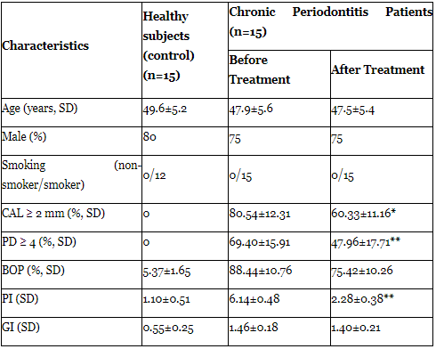
Thirty patients with pre-existing chronic severe periodontitis were included in the test group before and after conventional periodontal I therapy. The assessment of clinical parameters was carried out by a trained periodontist following criteria based on clinical parameters and the severity of periodontal tissue destruction [20]. The characteristic clinical signs of periodontitis included loss of clinical attachment, horizontal and/or angular alveolar bone loss, periodontal pocket formation, and gingival inflammation. To be included in the study, at least six sites with ongoing periodontal disease were required. Clinical measurements in patients with periodontitis included sites with alveolar bone loss>2mm and a pocket depth>5 mm with bleeding and attachment loss>3 mm. No subject (periodontal patient or healthy individual) had any systemic illness and they were all never-smokers. Patients with periodontitis had not received periodontal treatment or antibiotics within the preceding 5 months or any anti-inflammatory drug within 3 weeks prior to the study. The clinical characteristics of the study population and the healthy subjects (controls) are shown in (Table 1).
The control group consisted of thirty healthy non-smoker volunter males of the same age range, and with a clinically healthy periodontium from the same department. In the healthy subjects (control group), the probing depth was<3 mm and the attachment loss was<2 mm. Moreover, probing pocket depth and clinical attachment level were assessed at six sites per tooth and bleeding on probing at four sites per tooth. This group had<10% of sites with bleeding on probing (BOP), no sites with probing depth (PD)≥4 mm, no clinical attachment loss (AL),<2 mm, and no radiographic evidence of bone loss.
Exclusion criteria were individuals who had undergone periodontal treatment in the last 6 months; history of medications including antibiotics and anti-inflammatory drugs in the last 6 months; history of any systemic disease; and detection of any obvious oral mucosal lesion.
This study was approved by the Ethical Commission of the School of Dentistry, University of Buenos Aires. Written informed consent was obtained from each participant in this study, which was conducted under the rules of the Helsinki Declaration.
Saliva collection
Participants were instructed to refrain from eating, drinking and practicing oral hygiene procedures 12 hours before saliva collection. Whole unstimulated saliva was collected early morning after fasting (8 hours) from all patients by expectoration into sterile bulbs. The total amount of saliva collected was 1 ml. Collected samples were placed immediately on ice and transported to the laboratory, where they were centrifuged at 5,000 g for 10 minutes, and the clear supernatants were stored in aliquots at –80ºC in presence of protease inhibitors. The samples were thawed and the assays were performed with in 2 months of collection.
Clinical parameters
Clinical data included: CAL: clinical attachment level; PD: probing depth; BOP: bleeding on probing; PI: periodontal index; GI: gingival index (Table 1). The index measurements were done at six sites per tooth (mesio-buccal, mid-buccal, disto-buccal, disto-lingual, mid-lingual and mesio-lingual) and CAL was obtained by measuring only interproximal sites. These data were recorded and the treatment was performed by a trained periodontist. The data were recorded after saliva sample collection.
Periodontal therapy
Periodontal phase I therapy (PPIT) consisted of full-mouth scaling, root planning and oral hygiene instructions. Only the patients with periodontitis received PPIT after saliva sample collection and clinical measurements of baseline. The therapy was completed in two visits, each one week apart. No antibiotics were prescribed after the treatment. The patients were re-evaluated for clinical parameters one month afterwards and saliva samples were taken again.
Purification of saliva IgA
The IgA fractions of 15 patients with chronic periodontitis untreated and treated with PPIT and 15 healthy individuals were independently purified by standard diethylaminoethyl cellulose (DEAE) chromatography. Briefly, saliva samples were dialyzed against 0.01 M phosphate buffer, pH 8.0, for 18 hours and then applied to DEAE cellulose columns equilibrated in the same buffer. The pass-through IgG-rich fractions were discarded and IgA-rich fractions were eluted with 0.05 M NaCl in 0.01 M phosphate buffer, pH 8. The IgA concentration in the enriched fractions was quantificated by radial immunodiffusion method after concentration by ultrafiltration with PM-30 filtering membranes (Amicon, Beverly, MA, USA) (cutoff molecular weight, 30,000 Da). The concentration of IgA was 35.8±15.2 mg/dl in the IgA-enriched fractions. IgA was also purified by affinity chromatography of different saliva on Jacalin agarose beads following the recommendations of the supplier (ICN Pharmaceuticals, Irvine, CA, USA) and previously described methods [21].
ELISA procedure
Fifty microliters of synthetic β1 peptide solution (20 µg/ml) in 0.1 M Na2CO3 buffer, pH 9.6, was used to coat microtiter plates (NUNC, Kastrup, Denmark) at 4°C overnight.
After blocking the wells, diluted sera from patients with chronic severe periodontitis and healthy individuals (control) were added in triplicate and allowed to react with the peptide for 2 hours at 37°C. After the wells were thoroughly washed with 0.05% Tween 20 in phosphate buffered solution (PBS) 100 µl of 1:6000 biotinylated goat anti-human IgA antibodies (Sigma Chemical Co., St. Louis, MO, USA) was added and incubated for 1 hour at 37°C. Then, a 1:6000 dilution of extravidin-alkaline phosphatase (Sigma) was allowed to react an extra 30 minutes at 37°C. After extensive washings, p-nitrophenylphosphate (1 mg/ml) was added as the substrate, and the reaction was stopped at 30 minutes. Finally, the plates were read at 405 nm and the results for each sample were expressed as the means and SD of triplicate values.
Biomarker analysis
Concentration of salivary IL-1β (pg/ml), PGE2 (ng/ml), nitrites (µM) and MMP-3 (ng/ml) were determined in duplicate using ELISA assay for each patient and the healthy individuals of the human colorimetric immunoreactive kits from Cayman Chemical (Ann Arbor, MI, USA), respectively.
Drugs
The synthetic β1 peptide corresponded to the sequence of the second extracellular loop of the human β1-AR (H-W-W-RA- E-S-D-E-A-R-R-C-Y-N-D-P-K-C-C-D-F-V-T-N-R-C) and a 27-mer non-related peptide S-G-S-G-S-G-S-G-S-G-S-G-S-G-S-G-S-GS- G-S-G-S-G-S-G-S, as a negative control, were synthesized by Peptide Genetic Research Company (Livermore, CA, USA) as described previously [22].
Statistical analysis
Student's t-test for unpaired values was used to determine the levels of significance. Analysis of variance (ANOVA) and a post hoc test (Dunnett's method and Student-Newman-Keuls test) were employed when pair-wise multiple comparison procedures were necessary. Differences between means were considered significant at P<0.05.
It can be seen in (Table 2) the total salivary IgA (Total IgA (µg/ml) in periodontitis patients: 164±19; healthy subjects: 180±22; n=15 in each group) and its subtypes: IgA1 (µg/ ml): periodontitis patients: 66±5*; healthy subjects: 72± 6; *P<0.0001 comparing with healthy subjects; n=15 in each group and IgA2 (µg/ml): periodontitis patients: 98±8*; healthy subjects: 108±9; *P<0.0001 comparing with healthy subjects; n=15 in each group. Also, the values of the salivary flow (Basal salivary flow (ml/10 min) in periodontitis patients: 1.2±0.6*; healthy subjects: 9.7±3.5; *P<0.0001 comparing with healthy subjects; n=15 in each group) were performed with commercial plates for radial immunodiffusion containing anti-IgA.
The distribution of anti-β1-AR IgA was studied in clarified human whole saliva (CHWS) from untreated chronic periodontitis patients, treated with PPIT chronic severe periodontitis patients and in healthy individuals (control). The scattergram of (Figure 1) shows that the optical density values (at 405 nm) of salivary anti-β1-AR IgA from CHWS of the untreated chronic severe periodontitis patients (periodontal CHWS) and those from chronic severe periodontitis patients treated with PPIT. Also, the optical density values in healthy individuals (control CHWS) was shown. It can be seen that the data from periodontal CHWS and periodontal CHWS+PPIT were significantly higher (P<0.0001) than those of control CHWS. It is important to note that there is no significant difference between patients with chronic periodontitis before and after PPIT in the amount of this autoantibody. On the other hand, the immunoreactivity of saliva fron untrated and treated patients were negative in the presence of non-related peptide (data not shown), asserting the immunological recognition of the anti-β1 AR IgA salivary only when the coating antigen is the synthetic β1 peptide.
The fair amount is known about the immunological mechanisms responsible for the pathology observed in the disease. The morphology of chronic periodontitis lesions, and the clinical signs and symptoms of the disease suggest that cytokines (IL-1β, PGE2) [23,24], nitric oxide levels [25] and cytoplasmic enzymes (MMP-3) [26] are important in the pathogenesis of the disorders. But, we considerer important to determine if the anti β1 adrenoceptor IgA present in the saliva of chronic severe patients, participates in the pathophysiology of the disease.
In this study we demonstrated high concentrations of IL-1β, PGE2, nitrites and MMP-3 in saliva from untreated chronic severe periodontitis patients. Moreover, the highest levels of IL-1β and PGE2 were found in the saliva of these patients compared with those detected in healthy individuals (control). Previously was demonstrated that in human gingival fibroblasts are able to produce large amounts of PGE2 in response to inflammatory cytokines, and the increased PGE2 would be a potent stimulator of bone resorption [27]. Macrophages, mononuclear cells and fibroblasts from gingival tissues and endothelial cells are responsible for the increase in IL-1β production [28]; thus, there is a close association between IL-1β levels and periodontal disease status. After periodontal based therapy in patients with chronic periodontitis, IL-1β levels are reduced in all patients tested, which is correlated with clinical improvement. PGE2 is thought to be involved in the pathogenesis of the oral lesions observed in untreated chronic periodontitis, because of its role as a potent stimulator of bone resorption and association with attachment loss was published [29]. Therefore, there is a reciprocal interaction between PGE2 and IL-1β; IL-1β is a potent stimulator of PGE2 production in human gingival fibroblasts. However, PGE2 differential regulates IL-1β-induced matrix metalloproteinase (MMP-3) production. In human gingival fibroblasts from healthy gingiva, PGE2 down-regulates IL-1β-induced MMP-3 production, whereas in human gingival fibroblasts from periodontitis patients, PGE2 enhances it [29]. These data may reflect the heterogeneity of immuno-inflammatory responses in healthy and disease conditions, in which the concentrations of IL-1β, PGE2 MMP [30-31] may play a critical role as a marker of chronic severe periodontitis disease progression and oral manifestations.
The correlation between the amount of PGE2 and MMP-3 in the saliva of each patient studied in this study has demonstrated an important relationship with the amount of anti-β1 IgA. Analysis of this result, PGE2 and MMP-3 performer on follow up studies underlines the correlation on the levels of the cytokine and the enzyme in the saliva of untreated patients with chronic periodontitis and the presence of salivary anti-β1 IgA. It is important to note that, as previously reported, the pathogenesis of periodontal disease involves essential immunologic factors associated with infections caused by bacteria in sub-gingival plaques. The level of nitrite in saliva and its increment in patients with untreated chronic periodontitis was observed, and also an increased expression of iNOS in periodontal disease biopsy samples as well as in gingival fibroblast cell culture was described [22,32]. NO levels are associated with the severity of periodontitis, allowing differentiation between moderate and advanced generalized chronic periodontitis and NO levels were correlated with probing depth [25].
The biological plausibility of the differences observed in this study indicated that nitric oxide levels may be important in the pathogenesis of the disorders, and may be only in partly explained, by periodontal bacterial components triggering the host-immune response and causing inflammation and activation of pro-inflammatory mediators (IL-1β, PGE2 and MMP-3). All of these molecules travelling in blood, together with those produced locally by the inflammatory process in the soft and hard oral tissues, might influence the pathophysiology of chronic periodontitis, but the real importance of the presence in the saliva of an anti β1 adrenoceptor IgA remain to be determined.
The findings of the markedly elevated salivary IL-1β, PGE2 and MMP-3 levels and their significant reduction post therapy in patients with chronic periodontitis compared to healthy individuals (control), suggest a close association between salivary cytokines and enzymes and the periodontal status. But, the real participation of an anti β1adrenoceptor IgA present in the saliva of patients with severe chronic periodontitis, incorporated another pathophysiological factor in this multifactorial disease was not clarified yet in this present work. Future longitudinal studies with larger sample sizes are needed to validate in saliva not only if IL-1β, PGE2 and MMP-3 are "real biomarkers" for periodontal disease but to know what role would play the autoantibody (β1 adrenoceptor IgA) present in the saliva of these patients in the course of the periodontal disease or in its pathophysiology.
Clearly Auctoresonline and particularly Psychology and Mental Health Care Journal is dedicated to improving health care services for individuals and populations. The editorial boards' ability to efficiently recognize and share the global importance of health literacy with a variety of stakeholders. Auctoresonline publishing platform can be used to facilitate of optimal client-based services and should be added to health care professionals' repertoire of evidence-based health care resources.

Journal of Clinical Cardiology and Cardiovascular Intervention The submission and review process was adequate. However I think that the publication total value should have been enlightened in early fases. Thank you for all.

Journal of Women Health Care and Issues By the present mail, I want to say thank to you and tour colleagues for facilitating my published article. Specially thank you for the peer review process, support from the editorial office. I appreciate positively the quality of your journal.

Journal of Clinical Research and Reports I would be very delighted to submit my testimonial regarding the reviewer board and the editorial office. The reviewer board were accurate and helpful regarding any modifications for my manuscript. And the editorial office were very helpful and supportive in contacting and monitoring with any update and offering help. It was my pleasure to contribute with your promising Journal and I am looking forward for more collaboration.

We would like to thank the Journal of Thoracic Disease and Cardiothoracic Surgery because of the services they provided us for our articles. The peer-review process was done in a very excellent time manner, and the opinions of the reviewers helped us to improve our manuscript further. The editorial office had an outstanding correspondence with us and guided us in many ways. During a hard time of the pandemic that is affecting every one of us tremendously, the editorial office helped us make everything easier for publishing scientific work. Hope for a more scientific relationship with your Journal.

The peer-review process which consisted high quality queries on the paper. I did answer six reviewers’ questions and comments before the paper was accepted. The support from the editorial office is excellent.

Journal of Neuroscience and Neurological Surgery. I had the experience of publishing a research article recently. The whole process was simple from submission to publication. The reviewers made specific and valuable recommendations and corrections that improved the quality of my publication. I strongly recommend this Journal.

Dr. Katarzyna Byczkowska My testimonial covering: "The peer review process is quick and effective. The support from the editorial office is very professional and friendly. Quality of the Clinical Cardiology and Cardiovascular Interventions is scientific and publishes ground-breaking research on cardiology that is useful for other professionals in the field.

Thank you most sincerely, with regard to the support you have given in relation to the reviewing process and the processing of my article entitled "Large Cell Neuroendocrine Carcinoma of The Prostate Gland: A Review and Update" for publication in your esteemed Journal, Journal of Cancer Research and Cellular Therapeutics". The editorial team has been very supportive.

Testimony of Journal of Clinical Otorhinolaryngology: work with your Reviews has been a educational and constructive experience. The editorial office were very helpful and supportive. It was a pleasure to contribute to your Journal.

Dr. Bernard Terkimbi Utoo, I am happy to publish my scientific work in Journal of Women Health Care and Issues (JWHCI). The manuscript submission was seamless and peer review process was top notch. I was amazed that 4 reviewers worked on the manuscript which made it a highly technical, standard and excellent quality paper. I appreciate the format and consideration for the APC as well as the speed of publication. It is my pleasure to continue with this scientific relationship with the esteem JWHCI.

This is an acknowledgment for peer reviewers, editorial board of Journal of Clinical Research and Reports. They show a lot of consideration for us as publishers for our research article “Evaluation of the different factors associated with side effects of COVID-19 vaccination on medical students, Mutah university, Al-Karak, Jordan”, in a very professional and easy way. This journal is one of outstanding medical journal.

Dear Hao Jiang, to Journal of Nutrition and Food Processing We greatly appreciate the efficient, professional and rapid processing of our paper by your team. If there is anything else we should do, please do not hesitate to let us know. On behalf of my co-authors, we would like to express our great appreciation to editor and reviewers.

As an author who has recently published in the journal "Brain and Neurological Disorders". I am delighted to provide a testimonial on the peer review process, editorial office support, and the overall quality of the journal. The peer review process at Brain and Neurological Disorders is rigorous and meticulous, ensuring that only high-quality, evidence-based research is published. The reviewers are experts in their fields, and their comments and suggestions were constructive and helped improve the quality of my manuscript. The review process was timely and efficient, with clear communication from the editorial office at each stage. The support from the editorial office was exceptional throughout the entire process. The editorial staff was responsive, professional, and always willing to help. They provided valuable guidance on formatting, structure, and ethical considerations, making the submission process seamless. Moreover, they kept me informed about the status of my manuscript and provided timely updates, which made the process less stressful. The journal Brain and Neurological Disorders is of the highest quality, with a strong focus on publishing cutting-edge research in the field of neurology. The articles published in this journal are well-researched, rigorously peer-reviewed, and written by experts in the field. The journal maintains high standards, ensuring that readers are provided with the most up-to-date and reliable information on brain and neurological disorders. In conclusion, I had a wonderful experience publishing in Brain and Neurological Disorders. The peer review process was thorough, the editorial office provided exceptional support, and the journal's quality is second to none. I would highly recommend this journal to any researcher working in the field of neurology and brain disorders.
Dear Agrippa Hilda, Journal of Neuroscience and Neurological Surgery, Editorial Coordinator, I trust this message finds you well. I want to extend my appreciation for considering my article for publication in your esteemed journal. I am pleased to provide a testimonial regarding the peer review process and the support received from your editorial office. The peer review process for my paper was carried out in a highly professional and thorough manner. The feedback and comments provided by the authors were constructive and very useful in improving the quality of the manuscript. This rigorous assessment process undoubtedly contributes to the high standards maintained by your journal.

International Journal of Clinical Case Reports and Reviews. I strongly recommend to consider submitting your work to this high-quality journal. The support and availability of the Editorial staff is outstanding and the review process was both efficient and rigorous.

Thank you very much for publishing my Research Article titled “Comparing Treatment Outcome Of Allergic Rhinitis Patients After Using Fluticasone Nasal Spray And Nasal Douching" in the Journal of Clinical Otorhinolaryngology. As Medical Professionals we are immensely benefited from study of various informative Articles and Papers published in this high quality Journal. I look forward to enriching my knowledge by regular study of the Journal and contribute my future work in the field of ENT through the Journal for use by the medical fraternity. The support from the Editorial office was excellent and very prompt. I also welcome the comments received from the readers of my Research Article.

Dear Erica Kelsey, Editorial Coordinator of Cancer Research and Cellular Therapeutics Our team is very satisfied with the processing of our paper by your journal. That was fast, efficient, rigorous, but without unnecessary complications. We appreciated the very short time between the submission of the paper and its publication on line on your site.

I am very glad to say that the peer review process is very successful and fast and support from the Editorial Office. Therefore, I would like to continue our scientific relationship for a long time. And I especially thank you for your kindly attention towards my article. Have a good day!

"We recently published an article entitled “Influence of beta-Cyclodextrins upon the Degradation of Carbofuran Derivatives under Alkaline Conditions" in the Journal of “Pesticides and Biofertilizers” to show that the cyclodextrins protect the carbamates increasing their half-life time in the presence of basic conditions This will be very helpful to understand carbofuran behaviour in the analytical, agro-environmental and food areas. We greatly appreciated the interaction with the editor and the editorial team; we were particularly well accompanied during the course of the revision process, since all various steps towards publication were short and without delay".

I would like to express my gratitude towards you process of article review and submission. I found this to be very fair and expedient. Your follow up has been excellent. I have many publications in national and international journal and your process has been one of the best so far. Keep up the great work.

We are grateful for this opportunity to provide a glowing recommendation to the Journal of Psychiatry and Psychotherapy. We found that the editorial team were very supportive, helpful, kept us abreast of timelines and over all very professional in nature. The peer review process was rigorous, efficient and constructive that really enhanced our article submission. The experience with this journal remains one of our best ever and we look forward to providing future submissions in the near future.

I am very pleased to serve as EBM of the journal, I hope many years of my experience in stem cells can help the journal from one way or another. As we know, stem cells hold great potential for regenerative medicine, which are mostly used to promote the repair response of diseased, dysfunctional or injured tissue using stem cells or their derivatives. I think Stem Cell Research and Therapeutics International is a great platform to publish and share the understanding towards the biology and translational or clinical application of stem cells.

I would like to give my testimony in the support I have got by the peer review process and to support the editorial office where they were of asset to support young author like me to be encouraged to publish their work in your respected journal and globalize and share knowledge across the globe. I really give my great gratitude to your journal and the peer review including the editorial office.

I am delighted to publish our manuscript entitled "A Perspective on Cocaine Induced Stroke - Its Mechanisms and Management" in the Journal of Neuroscience and Neurological Surgery. The peer review process, support from the editorial office, and quality of the journal are excellent. The manuscripts published are of high quality and of excellent scientific value. I recommend this journal very much to colleagues.

Dr.Tania Muñoz, My experience as researcher and author of a review article in The Journal Clinical Cardiology and Interventions has been very enriching and stimulating. The editorial team is excellent, performs its work with absolute responsibility and delivery. They are proactive, dynamic and receptive to all proposals. Supporting at all times the vast universe of authors who choose them as an option for publication. The team of review specialists, members of the editorial board, are brilliant professionals, with remarkable performance in medical research and scientific methodology. Together they form a frontline team that consolidates the JCCI as a magnificent option for the publication and review of high-level medical articles and broad collective interest. I am honored to be able to share my review article and open to receive all your comments.

“The peer review process of JPMHC is quick and effective. Authors are benefited by good and professional reviewers with huge experience in the field of psychology and mental health. The support from the editorial office is very professional. People to contact to are friendly and happy to help and assist any query authors might have. Quality of the Journal is scientific and publishes ground-breaking research on mental health that is useful for other professionals in the field”.

Dear editorial department: On behalf of our team, I hereby certify the reliability and superiority of the International Journal of Clinical Case Reports and Reviews in the peer review process, editorial support, and journal quality. Firstly, the peer review process of the International Journal of Clinical Case Reports and Reviews is rigorous, fair, transparent, fast, and of high quality. The editorial department invites experts from relevant fields as anonymous reviewers to review all submitted manuscripts. These experts have rich academic backgrounds and experience, and can accurately evaluate the academic quality, originality, and suitability of manuscripts. The editorial department is committed to ensuring the rigor of the peer review process, while also making every effort to ensure a fast review cycle to meet the needs of authors and the academic community. Secondly, the editorial team of the International Journal of Clinical Case Reports and Reviews is composed of a group of senior scholars and professionals with rich experience and professional knowledge in related fields. The editorial department is committed to assisting authors in improving their manuscripts, ensuring their academic accuracy, clarity, and completeness. Editors actively collaborate with authors, providing useful suggestions and feedback to promote the improvement and development of the manuscript. We believe that the support of the editorial department is one of the key factors in ensuring the quality of the journal. Finally, the International Journal of Clinical Case Reports and Reviews is renowned for its high- quality articles and strict academic standards. The editorial department is committed to publishing innovative and academically valuable research results to promote the development and progress of related fields. The International Journal of Clinical Case Reports and Reviews is reasonably priced and ensures excellent service and quality ratio, allowing authors to obtain high-level academic publishing opportunities in an affordable manner. I hereby solemnly declare that the International Journal of Clinical Case Reports and Reviews has a high level of credibility and superiority in terms of peer review process, editorial support, reasonable fees, and journal quality. Sincerely, Rui Tao.

Clinical Cardiology and Cardiovascular Interventions I testity the covering of the peer review process, support from the editorial office, and quality of the journal.

Clinical Cardiology and Cardiovascular Interventions, we deeply appreciate the interest shown in our work and its publication. It has been a true pleasure to collaborate with you. The peer review process, as well as the support provided by the editorial office, have been exceptional, and the quality of the journal is very high, which was a determining factor in our decision to publish with you.

The peer reviewers process is quick and effective, the supports from editorial office is excellent, the quality of journal is high. I would like to collabroate with Internatioanl journal of Clinical Case Reports and Reviews journal clinically in the future time.

Clinical Cardiology and Cardiovascular Interventions, I would like to express my sincerest gratitude for the trust placed in our team for the publication in your journal. It has been a true pleasure to collaborate with you on this project. I am pleased to inform you that both the peer review process and the attention from the editorial coordination have been excellent. Your team has worked with dedication and professionalism to ensure that your publication meets the highest standards of quality. We are confident that this collaboration will result in mutual success, and we are eager to see the fruits of this shared effort.

Dear Dr. Jessica Magne, Editorial Coordinator 0f Clinical Cardiology and Cardiovascular Interventions, I hope this message finds you well. I want to express my utmost gratitude for your excellent work and for the dedication and speed in the publication process of my article titled "Navigating Innovation: Qualitative Insights on Using Technology for Health Education in Acute Coronary Syndrome Patients." I am very satisfied with the peer review process, the support from the editorial office, and the quality of the journal. I hope we can maintain our scientific relationship in the long term.

Dear Monica Gissare, - Editorial Coordinator of Nutrition and Food Processing. ¨My testimony with you is truly professional, with a positive response regarding the follow-up of the article and its review, you took into account my qualities and the importance of the topic¨.

Dear Dr. Jessica Magne, Editorial Coordinator 0f Clinical Cardiology and Cardiovascular Interventions, The review process for the article “The Handling of Anti-aggregants and Anticoagulants in the Oncologic Heart Patient Submitted to Surgery” was extremely rigorous and detailed. From the initial submission to the final acceptance, the editorial team at the “Journal of Clinical Cardiology and Cardiovascular Interventions” demonstrated a high level of professionalism and dedication. The reviewers provided constructive and detailed feedback, which was essential for improving the quality of our work. Communication was always clear and efficient, ensuring that all our questions were promptly addressed. The quality of the “Journal of Clinical Cardiology and Cardiovascular Interventions” is undeniable. It is a peer-reviewed, open-access publication dedicated exclusively to disseminating high-quality research in the field of clinical cardiology and cardiovascular interventions. The journal's impact factor is currently under evaluation, and it is indexed in reputable databases, which further reinforces its credibility and relevance in the scientific field. I highly recommend this journal to researchers looking for a reputable platform to publish their studies.

Dear Editorial Coordinator of the Journal of Nutrition and Food Processing! "I would like to thank the Journal of Nutrition and Food Processing for including and publishing my article. The peer review process was very quick, movement and precise. The Editorial Board has done an extremely conscientious job with much help, valuable comments and advices. I find the journal very valuable from a professional point of view, thank you very much for allowing me to be part of it and I would like to participate in the future!”

Dealing with The Journal of Neurology and Neurological Surgery was very smooth and comprehensive. The office staff took time to address my needs and the response from editors and the office was prompt and fair. I certainly hope to publish with this journal again.Their professionalism is apparent and more than satisfactory. Susan Weiner

My Testimonial Covering as fellowing: Lin-Show Chin. The peer reviewers process is quick and effective, the supports from editorial office is excellent, the quality of journal is high. I would like to collabroate with Internatioanl journal of Clinical Case Reports and Reviews.

My experience publishing in Psychology and Mental Health Care was exceptional. The peer review process was rigorous and constructive, with reviewers providing valuable insights that helped enhance the quality of our work. The editorial team was highly supportive and responsive, making the submission process smooth and efficient. The journal's commitment to high standards and academic rigor makes it a respected platform for quality research. I am grateful for the opportunity to publish in such a reputable journal.

My experience publishing in International Journal of Clinical Case Reports and Reviews was exceptional. I Come forth to Provide a Testimonial Covering the Peer Review Process and the editorial office for the Professional and Impartial Evaluation of the Manuscript.

I would like to offer my testimony in the support. I have received through the peer review process and support the editorial office where they are to support young authors like me, encourage them to publish their work in your esteemed journals, and globalize and share knowledge globally. I really appreciate your journal, peer review, and editorial office.

Dear Agrippa Hilda- Editorial Coordinator of Journal of Neuroscience and Neurological Surgery, "The peer review process was very quick and of high quality, which can also be seen in the articles in the journal. The collaboration with the editorial office was very good."

I would like to express my sincere gratitude for the support and efficiency provided by the editorial office throughout the publication process of my article, “Delayed Vulvar Metastases from Rectal Carcinoma: A Case Report.” I greatly appreciate the assistance and guidance I received from your team, which made the entire process smooth and efficient. The peer review process was thorough and constructive, contributing to the overall quality of the final article. I am very grateful for the high level of professionalism and commitment shown by the editorial staff, and I look forward to maintaining a long-term collaboration with the International Journal of Clinical Case Reports and Reviews.

To Dear Erin Aust, I would like to express my heartfelt appreciation for the opportunity to have my work published in this esteemed journal. The entire publication process was smooth and well-organized, and I am extremely satisfied with the final result. The Editorial Team demonstrated the utmost professionalism, providing prompt and insightful feedback throughout the review process. Their clear communication and constructive suggestions were invaluable in enhancing my manuscript, and their meticulous attention to detail and dedication to quality are truly commendable. Additionally, the support from the Editorial Office was exceptional. From the initial submission to the final publication, I was guided through every step of the process with great care and professionalism. The team's responsiveness and assistance made the entire experience both easy and stress-free. I am also deeply impressed by the quality and reputation of the journal. It is an honor to have my research featured in such a respected publication, and I am confident that it will make a meaningful contribution to the field.

"I am grateful for the opportunity of contributing to [International Journal of Clinical Case Reports and Reviews] and for the rigorous review process that enhances the quality of research published in your esteemed journal. I sincerely appreciate the time and effort of your team who have dedicatedly helped me in improvising changes and modifying my manuscript. The insightful comments and constructive feedback provided have been invaluable in refining and strengthening my work".

I thank the ‘Journal of Clinical Research and Reports’ for accepting this article for publication. This is a rigorously peer reviewed journal which is on all major global scientific data bases. I note the review process was prompt, thorough and professionally critical. It gave us an insight into a number of important scientific/statistical issues. The review prompted us to review the relevant literature again and look at the limitations of the study. The peer reviewers were open, clear in the instructions and the editorial team was very prompt in their communication. This journal certainly publishes quality research articles. I would recommend the journal for any future publications.

Dear Jessica Magne, with gratitude for the joint work. Fast process of receiving and processing the submitted scientific materials in “Clinical Cardiology and Cardiovascular Interventions”. High level of competence of the editors with clear and correct recommendations and ideas for enriching the article.

We found the peer review process quick and positive in its input. The support from the editorial officer has been very agile, always with the intention of improving the article and taking into account our subsequent corrections.

My article, titled 'No Way Out of the Smartphone Epidemic Without Considering the Insights of Brain Research,' has been republished in the International Journal of Clinical Case Reports and Reviews. The review process was seamless and professional, with the editors being both friendly and supportive. I am deeply grateful for their efforts.

To Dear Erin Aust – Editorial Coordinator of Journal of General Medicine and Clinical Practice! I declare that I am absolutely satisfied with your work carried out with great competence in following the manuscript during the various stages from its receipt, during the revision process to the final acceptance for publication. Thank Prof. Elvira Farina

Dear Jessica, and the super professional team of the ‘Clinical Cardiology and Cardiovascular Interventions’ I am sincerely grateful to the coordinated work of the journal team for the no problem with the submission of my manuscript: “Cardiometabolic Disorders in A Pregnant Woman with Severe Preeclampsia on the Background of Morbid Obesity (Case Report).” The review process by 5 experts was fast, and the comments were professional, which made it more specific and academic, and the process of publication and presentation of the article was excellent. I recommend that my colleagues publish articles in this journal, and I am interested in further scientific cooperation. Sincerely and best wishes, Dr. Oleg Golyanovskiy.

Dear Ashley Rosa, Editorial Coordinator of the journal - Psychology and Mental Health Care. " The process of obtaining publication of my article in the Psychology and Mental Health Journal was positive in all areas. The peer review process resulted in a number of valuable comments, the editorial process was collaborative and timely, and the quality of this journal has been quickly noticed, resulting in alternative journals contacting me to publish with them." Warm regards, Susan Anne Smith, PhD. Australian Breastfeeding Association.

Dear Jessica Magne, Editorial Coordinator, Clinical Cardiology and Cardiovascular Interventions, Auctores Publishing LLC. I appreciate the journal (JCCI) editorial office support, the entire team leads were always ready to help, not only on technical front but also on thorough process. Also, I should thank dear reviewers’ attention to detail and creative approach to teach me and bring new insights by their comments. Surely, more discussions and introduction of other hemodynamic devices would provide better prevention and management of shock states. Your efforts and dedication in presenting educational materials in this journal are commendable. Best wishes from, Farahnaz Fallahian.

Dear Maria Emerson, Editorial Coordinator, International Journal of Clinical Case Reports and Reviews, Auctores Publishing LLC. I am delighted to have published our manuscript, "Acute Colonic Pseudo-Obstruction (ACPO): A rare but serious complication following caesarean section." I want to thank the editorial team, especially Maria Emerson, for their prompt review of the manuscript, quick responses to queries, and overall support. Yours sincerely Dr. Victor Olagundoye.

Dear Ashley Rosa, Editorial Coordinator, International Journal of Clinical Case Reports and Reviews. Many thanks for publishing this manuscript after I lost confidence the editors were most helpful, more than other journals Best wishes from, Susan Anne Smith, PhD. Australian Breastfeeding Association.

Dear Agrippa Hilda, Editorial Coordinator, Journal of Neuroscience and Neurological Surgery. The entire process including article submission, review, revision, and publication was extremely easy. The journal editor was prompt and helpful, and the reviewers contributed to the quality of the paper. Thank you so much! Eric Nussbaum, MD

Dr Hala Al Shaikh This is to acknowledge that the peer review process for the article ’ A Novel Gnrh1 Gene Mutation in Four Omani Male Siblings, Presentation and Management ’ sent to the International Journal of Clinical Case Reports and Reviews was quick and smooth. The editorial office was prompt with easy communication.

Dear Erin Aust, Editorial Coordinator, Journal of General Medicine and Clinical Practice. We are pleased to share our experience with the “Journal of General Medicine and Clinical Practice”, following the successful publication of our article. The peer review process was thorough and constructive, helping to improve the clarity and quality of the manuscript. We are especially thankful to Ms. Erin Aust, the Editorial Coordinator, for her prompt communication and continuous support throughout the process. Her professionalism ensured a smooth and efficient publication experience. The journal upholds high editorial standards, and we highly recommend it to fellow researchers seeking a credible platform for their work. Best wishes By, Dr. Rakhi Mishra.

Dear Jessica Magne, Editorial Coordinator, Clinical Cardiology and Cardiovascular Interventions, Auctores Publishing LLC. The peer review process of the journal of Clinical Cardiology and Cardiovascular Interventions was excellent and fast, as was the support of the editorial office and the quality of the journal. Kind regards Walter F. Riesen Prof. Dr. Dr. h.c. Walter F. Riesen.

Dear Ashley Rosa, Editorial Coordinator, International Journal of Clinical Case Reports and Reviews, Auctores Publishing LLC. Thank you for publishing our article, Exploring Clozapine's Efficacy in Managing Aggression: A Multiple Single-Case Study in Forensic Psychiatry in the international journal of clinical case reports and reviews. We found the peer review process very professional and efficient. The comments were constructive, and the whole process was efficient. On behalf of the co-authors, I would like to thank you for publishing this article. With regards, Dr. Jelle R. Lettinga.

Dear Clarissa Eric, Editorial Coordinator, Journal of Clinical Case Reports and Studies, I would like to express my deep admiration for the exceptional professionalism demonstrated by your journal. I am thoroughly impressed by the speed of the editorial process, the substantive and insightful reviews, and the meticulous preparation of the manuscript for publication. Additionally, I greatly appreciate the courteous and immediate responses from your editorial office to all my inquiries. Best Regards, Dariusz Ziora

Dear Chrystine Mejia, Editorial Coordinator, Journal of Neurodegeneration and Neurorehabilitation, Auctores Publishing LLC, We would like to thank the editorial team for the smooth and high-quality communication leading up to the publication of our article in the Journal of Neurodegeneration and Neurorehabilitation. The reviewers have extensive knowledge in the field, and their relevant questions helped to add value to our publication. Kind regards, Dr. Ravi Shrivastava.

Dear Clarissa Eric, Editorial Coordinator, Journal of Clinical Case Reports and Studies, Auctores Publishing LLC, USA Office: +1-(302)-520-2644. I would like to express my sincere appreciation for the efficient and professional handling of my case report by the ‘Journal of Clinical Case Reports and Studies’. The peer review process was not only fast but also highly constructive—the reviewers’ comments were clear, relevant, and greatly helped me improve the quality and clarity of my manuscript. I also received excellent support from the editorial office throughout the process. Communication was smooth and timely, and I felt well guided at every stage, from submission to publication. The overall quality and rigor of the journal are truly commendable. I am pleased to have published my work with Journal of Clinical Case Reports and Studies, and I look forward to future opportunities for collaboration. Sincerely, Aline Tollet, UCLouvain.

Dear Ms. Mayra Duenas, Editorial Coordinator, International Journal of Clinical Case Reports and Reviews. “The International Journal of Clinical Case Reports and Reviews represented the “ideal house” to share with the research community a first experience with the use of the Simeox device for speech rehabilitation. High scientific reputation and attractive website communication were first determinants for the selection of this Journal, and the following submission process exceeded expectations: fast but highly professional peer review, great support by the editorial office, elegant graphic layout. Exactly what a dynamic research team - also composed by allied professionals - needs!" From, Chiara Beccaluva, PT - Italy.

Dear Maria Emerson, Editorial Coordinator, we have deeply appreciated the professionalism demonstrated by the International Journal of Clinical Case Reports and Reviews. The reviewers have extensive knowledge of our field and have been very efficient and fast in supporting the process. I am really looking forward to further collaboration. Thanks. Best regards, Dr. Claudio Ligresti

Dear Chrystine Mejia, Editorial Coordinator, Journal of Neurodegeneration and Neurorehabilitation. “The peer review process was efficient and constructive, and the editorial office provided excellent communication and support throughout. The journal ensures scientific rigor and high editorial standards, while also offering a smooth and timely publication process. We sincerely appreciate the work of the editorial team in facilitating the dissemination of innovative approaches such as the Bonori Method.” Best regards, Dr. Matteo Bonori.
