AUCTORES
Globalize your Research

Research Article | DOI: https://doi.org/10.31579/2690-8808/176
*Corresponding Author: Atul Khare, Department of surgery, Government Medical College Shadol, India.
Citation: Atul Khare, (2023), Duodenal Atresia- Clinical Presentation and Management in Tertiary Care Centre, J, Clinical Case Reports and Studies, 4(3); DOI:10.31579/2690-8808/176
Copyright: ©, 2023, Atul Khare. This is an open access article distributed under the Creative Commons Attribution License, which permits unrestricted use, distribution, and reproduction in any medium, provided the original work is properly cited.
Abstract
Introduction: Congenital duodenal obstruction (CDO) is a common surgical anomaly in newborns, that can be diagnosed prenatally and requires careful planning for surgical repair after birth. This study focuses on the surgical management of duodenal atres
Received: 28 August 2023 | Accepted: 04 September 2023 | Published: 11 September 2023
Keywords: ileal atresia; exploratory laparotomy; neonatal intestinal obstruction
Introduction: Congenital duodenal obstruction (CDO) is a common surgical anomaly in newborns, that can be diagnosed prenatally and requires careful planning for surgical repair after birth. This study focuses on the surgical management of duodenal atresia. Intestinal atresia is one of the most common and leading causes of neonatal intestinal obstruction (NIO), and second most common cause of NIO in many developing countries. In this study, we analysed the short term outcome of surgical management of intestinal atresia in our unit.
Aims and objectives: This prospective and retrospective cohort study done in one-year period from march 2021 to march 2022 in a tertiary care hospital in Indian population [ mainly west India]. The main aim of our study is- ‘’Duodenal atresia- clinical presentation and management in tertiary care centre’’.
Materials and methods: All patients of intestinal atresia admitted in our centre in neonatal unit in one-year period from march 2021 to march 2022 were included, the data recorded and analysed. Data of demographics, antenatal history, presentation, location, and type of IA (duodenal, jejuno-ileal, colonic), and peri-operative complications were recorded.
After making our presumptive diagnosis with clinical assessment, an upright X-ray abdomen was taken and Decision for surgery was finalised.
Statistical Package for Social Sciences (SPSS 15.0 version, SPSS Inc, Chicago Ill) was used for data entry and analysis. Results were expressed as means, ranges and percentages.
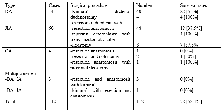
Results: 64 patients were male and 48 of them were female, M:F ratio was 1.3:1. Average weight of atresia was 2.14 kg [ranges from 1.1- 3.3kg] and mean age of presentation was 6.54 days [ranges from one day to 32 days]. Patients having age more than one week were only 28 [25%] in number. Cases were highest in December and lowest in the month of April. Muslim population were mostly associated with duodenal atresia. Intestinal atresia included different variants as follows, duodenal atresia- [n=44], Jejuno-ileal- [n=60], Colonic- [n=4], Multiple atresia- [n=4]. In duodenal atresia the distribution was as follows, DA- type-1 [n=14] in which annular pancreas [n=6] and perforated duodenal web [n=4], DA- type-2 [n=2] and DA-type-3 [n=28] cases. In duodenal atresia, TEF associated with [n=3], ARM associated with [n=3] and ARM with TEF both associated with [n=2] cases. In jejunoileal atresia type-1 [n=10], Type-2 [n=5], Type-3a [n=36], Type-3b [n=1], Type-4 [n=12] were recorded. In colonic atresia type-1 [n=3], type-2 [n=0] and type-3 [n=1] cases were recorded. In DA vomiting was the chief complaint while in JIA and IA abdominal distension, bilious vomiting and failure to pass meconium were the chief complains. All patients required surgical repair. A surprising result was found in jejunal atresia patients treated with tapering enteroplasty with trans-anastomotic tube with 100% survival rates. Most common complication was functional obstruction followed by SSI and anastomotic leak. Most of the patients required reoperation in JIA cases. Return of bowel function was seen in 4.3 days in DA, 6.2 days in JIA and 8.4 days in colonic atresia. Mean hospital stay for DA was noted to be 11.4 days, for jejunal atresia it was around 12.8 days and for CA it was found to be 11.5 days for survivors. Overall survival rates of surgery of intestinal atresia after surgery was 51.8%, among which DA accounted to 59.1%, jejuno-ileal atresia- 50% and for CA- 50%. During the one month follow up period, mainly cough and cold n=7 [6.25%], fever n=5 [4.46%], functional obstruction n=2 [1.78%] and with sepsis in n=1 [0.9%] were found.
Conclusions: Short term survival of neonates with intestinal atresias in our unit is still poor when compared with statistics from developed countries. Thus efforts are being made to improve the surgical outcome and to understand difficulties faced by surgeons with the help of this study.
Congenital duodenal obstruction (CDO) is a common surgical anomaly in newborns [1, 2] that can be diagnosed prenatally and requires careful planning for surgical repair after birth. This study focuses on the surgical management of duodenal atresia, a common form of CDO. With the rising popularity of laparoscopic surgery, duodenal atresia, one of the forms of CDO, can also be managed laparoscopically. However, use of a laparoscopic approach in these patients requires enormous experience in minimally invasive surgery along with supply of special equipments to perform such a technically challenging procedure in a limited operative space [3, 4].
Congenital duodenal obstruction accounts for 50% of all intestinal atresia cases [1, 2]. The incidence of the disease varies from 1 in 5000 to 1 in 10,000 newborns [5–8]. Limited information is available about hereditary forms of CDO. Unlike other types of congenital intestinal obstruction, duodenal obstruction has a high association with other anomalies; such concomitant anomalies are reported in 38% Atresia - Pathology and Therapeutic Approach of patients with CDO [9, 10]. The most common associated disease reported is Down syndrome registered in 25–46% of cases [11–13]. Other associated anomalies include intestinal malrotation (54%), congenital cardiac anomalies (32–48%), esophageal atresia (9%), renal anomalies (5%), and anorectal malformations (7%). These anomalies can be part of VACTERL syndrome or isolated [13]. 12% of patients with duodenal atresia have an associated anomaly of the biliary tract, such as biliary atresia [21, 22]. Associated diseases tend to determine the postoperative course in patients with duodenal atresia. In patients with associated esophageal atresia or cardiac defects—most often a complete atrioventricular septal defect—high mortality rates have been reported.
In most cases, the diagnosis of duodenal obstruction can be established prenatally. Duodenal obstruction develops approximately by 12–14 weeks of fetal development, so there is no possibility of earlier detection of this anomaly. Ultrasound is used to define the “double bubble sign.” These are two fluid levels, one in the distended stomach, and the other in the duodenum. Polyhydramnios develops in pregnancies complicated by duodenal obstruction. Postnatally, the diagnosis of duodenal obstruction is confirmed in an abdominal X-ray, showing the “double bubble” sign as described above. Abdominal ultrasound is necessary to detect not only duodenal atresia, but also to find concomitant anomalies and rare forms of situs inversus. These findings can necessitate alternative port placement during laparoscopy.
Currently, the standard method of recanalizing the duodenal lumen is a diamond-shaped duodenal anastomosis. The introduction of minimally invasive laparoscopic instruments, optical systems with small diameters, and high-resolution screens has expanded the potential of laparoscopy. These developments have increased the interest of paediatric surgeons in laparoscopy as a modality for reconstruction in patients with CDO. However, laparoscopic duodenal anastomosis is considered the most demanding surgical procedure in paediatric surgery. Therefore, this procedure is restricted to be performed in advanced centres specializing in minimally invasive surgery in neonates. Duodenal atresia is most often located in the second (descending) portion of the duodenum. Historically, such patients were treated surgically with laparotomy or laparoscopy. Despite the results of such treatment being satisfactory, these techniques are fraught with risks associated with the operation itself and general anaesthesia, and do not show good cosmetical results. In addition, the risk of adhesive intestinal obstruction in infants after laparotomy is approximately 6–14% and is absent if the peritoneum is left intact, as, for example, with transoral access.
Intestinal atresia is one of the most common and leading causes of neonatal intestinal obstruction (NIO), and second most common cause of NIO in many developing countries. [1‑3] mostly these patients, DA and JIA occur separately but sometimes they occur together in one patient.[4] In most developed countries surgical outcomes are improved due to various causes like availability of prenatal diagnosis, awareness in parents, early presentation of clinical features, availability of paediatric surgeons, availability of neonatal parenteral nutrition, neonatal anaesthesia, better postoperative care and neonatal surgical intensive care services.[5,6] In many low and middle income countries (LMICs), outcome has remained poor due to improper and less availability of these facility.[1,7]
It is a congenital obstruction of the intestine, sometimes associated with a loss of tissue, resulting in a disruption of intestinal continuity. The incidence of intestinal atresia is approximately 1 in 4000 live births. Etiopathogenesis of intestinal atresia is failure of recanalization of the initial solid-core phase of intestinal development, and in utero vascular accident is the cause of it hypothesized. This can occur anywhere in the intestinal tract from duodenum, jejunoileal region and colon. DA mostly associated with other congenital anomalies, most commonly down syndrome and associated with imperforated anus. Jejunoileal atresia’s occurs from Treitz ligament to ileocecal valve anywhere and mainly associated with cystic fibrosis and malrotation. Colonic atresias unusual in that they found in same anatomical region of colon [transverse colon] and with same degree of severity [lumen and mesentery loss. This is present with various degree of severity like mucosal web to complete lumen or mesentery loss and cause multiple atresia throughout the bowel.
In this study, we basically highlight the short‑term outcome of surgical management of intestinal atresia in our paediatric surgery unit.
This prospective and retrospective cohort study done in one-year period from march 2021 to march 2022 in a tertiary care hospital in Indian population [ mainly west India]. The main aims of our study are ‘’Duodenal atresia- clinical presentation and management in tertiary care centre’’.
All the patients of intestinal atresia admitted in our centre in neonatal unit in one-year period from march 2021 to march 2022 were included, their data recorded and analysed. All intestinal atresia patients selected and their history, demographic data, medical details, treatments, surgical outcome, hospital stay and complications were recorded. In detail we also recorded the data like antenatal history, presentation, location, and type of IA (duodenal, jejuno-ileal, colonic), and peri-operative complications.
Patients who died before the operation or before making any definitive diagnosis, with volvulus, complicated meconium ileus, gastroschisis and who left or got discharged against medical advice, were excluded.
After making our presumptive diagnosis with clinical assessment, an upright X-ray abdomen was taken and confirmed to have intestinal atresia. Patients were then vitally stabilised by intravenous fluid and antibiotics. Nasogastric (NG) tube was inserted in all patients. After making definitive diagnosis, plan for surgery was made. Postoperatively antibiotics were continued and NG tube was removed after peristalsis was re-established or when the NG aspirate was found to be gastric and < 15ml>
Statistical Package for Social Sciences (SPSS 15.0 version, SPSS Inc, Chicago Ill) was used for data entry and analysis. Results were expressed as means, ranges and percentages.
A total of 136 patients were studied, in which 6 patients expired before operation, 7 patients went LAMA, 5 patients came with previous history of operation, 5 patients came with atresia with meconium ileus, and 1 patient came with atresia associated with gastroschisis. These patients were excluded from the data, thus we analysed only 112 patients who required operation. We found these results from our study-
1] males were 64 and females were 48 patients, M/F ratio was 1.3:1.
2] average weight of atresia was 2.14 kg [ranges from 1.1- 3.3kg]
3] mean age of presentation was 6.54 days [ranges from one day to 32 days]. Patients having age more than one week were only 28 [25%].
4] monthly distribution of cases was highest in December and lowest in April months.

5] Muslim population was found to be mostly associated with duodenal atresia while Hindu population were mostly associated with all intestinal atresias.
6] We found that state wise distribution of cases from Rajasthan [n=95], Haryana [n=6], Madhya-Pradesh [n=1] and from Uttar-Pradesh [n=10] cases found
7] Intestinal atresia included duodenal atresia in [n=44], Jejuno-ileal in [n=60], Colonic in [n=4], Multiple atresias in [n=4].
8] In duodenal atresia DA- type-1 [n=14] in which annular pancreas [n=6] and perforated duodenal web [n=4], DA- type-2 [n=2] and DA-type- 3 [n=28] cases.

9] In duodenal atresia, trachea-oesophageal fistula associated with [n=3], anorectal malformation associated with [n=3] and anorectal malformation with trachea-oesophageal both associated with [n=2] cases.
10] in jejunileal atresia type-1 [n=10], Type-2 [n=5], Type-3a [n=36], Type-3b [n=1], Type-4 [n=12] are recorded.
11] in colonic atresia type-1 [n=3], type-2 [n=0] and type-3 [n=1] cases are recorded.
12] in DA vomiting is chief complain while in JIA and IA abdominal distension, bilious vomiting, and failure to pass meconium is chief complains.
13] in cases of DA; Double-bubble sign on X-ray abdomen was found, while in perforated duodenal web, distal gas with double-bubble was seen. In triple atresia patients, double-bubble sign with red rubber catheter in upper oesophageal pouch was found. In perforated duodenal web patient upper-GI gastrografin contrast study was performed because of diagnostic dilemma due to presence of gas in distal bowel on X-ray. In JIA, Tripple bubble sign, and multiple dilated bowel loops and multiple air-fluid levels were seen. Gastrografin enema was performed in cases of ileal atresia.

Multiple ileal atresia radiography= multiple bubble sign

Duodenal atresia= double bubble sign
14] Surrgical treatment was done in all cases after resuscitating the patient and proper investigations. A surprising result was found in jejunal atresia patients with anti-mesenteric tapering enteroplasty with trans-anastomotic tube showing 100% survival rates. Other procedures used were as follows-
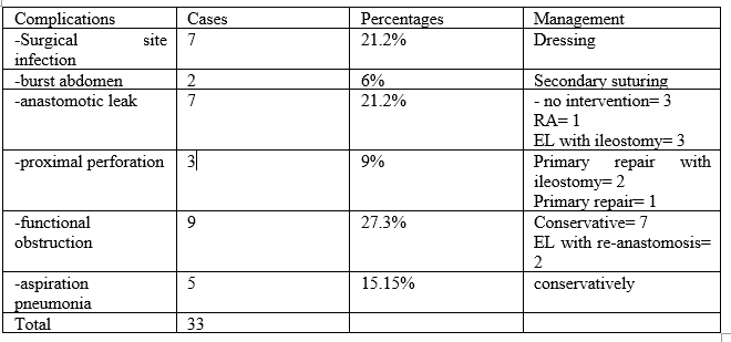
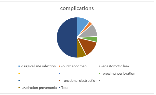
15] there were many postoperative complications. Most common complication being functional obstruction followed by SSI and anastomotic leak. Mostly reoperation was required in JIA cases.
16] return of bowel function was seen in 4.3 days in DA, 6.2 days in JIA and 8.4 days in colonic atresia.
17] mean hospital stay for duodenal atresia was 11.4 days, for jejunal atresia 12.8 days and for colonic atresia 11.5 days for survivors.
18] Overall survival rates of surgery of intestinal atresia after surgery was 51.8%. for duodenal atresia 59.1%, jejuno-ileal atresia- 50% and for colonic atresia- 50%.
19] one months follow up also taken in most operated patients which mainly showed cough and cold n=7 [6.25%], fever n=5 [4.46%], functional obstruction n=2 [1.78%] and with sepsis in n=1 [0.9%] .
Congenital duodenal obstruction (CDO) is a common surgical anomaly in newborns, that can be diagnosed prenatally and requires careful planning for surgical repair after birth. This chapter focuses on the surgical management of duodenal atresia, a common form of CDO. With the rising popularity of laparoscopic surgery, duodenal atresia, one of the forms of CDO, also can be managed laparoscopically. However, use of a laparoscopic approach in these patients requires advanced experience in minimally invasive surgery and special equipment to perform such a demanding procedure in a limited operative space.
IA is one of the most common causes of neonatal intestinal obstruction. In detection of duodenal atresia than JIA or CA, Prenatal ultrasonography is more reliable. Basu and Burge reported that 31% of patients with small bowel atresia could be diagnosed on prenatal ultrasound [11].
Pre-operative management was needed in all patients which included, primary resuscitation, correction of dehydration and electrolyte abnormalities., echocardiography and ultrasonography of the abdomen should be performed in all patients because of high incidence of cardiac and renal anomalies associated with DA [12]. In this study only n=10 [22.7%] DA patients associated with cardiac anomalies and only n=4 [9%] associated with imperforated anus. similarly in a series of 138 cases of DA, 38
We concluded that survival rates of neonates with intestinal atresia is poor in developing country, in a high volume tertiary care centre in India. It is not only because of surgical techniques and expertise but due to multifactorial causes like delayed presentation, high patients load at hospitals, overcrowding in the ICU and septicaemia. It leads to failure of short term survivals of patients. Late presentation is common in this series but does not appear to negatively affect the outcome as meticulous pre‑operative resuscitation is emphasized. A high proportion of the mortalities were seen in cases with re‑operation for anastomotic leak.
From this study we found that we can improve surgical outcomes by improving prenatal diagnosis, early referral to higher centre, planned delivery in centres, establishment of neonatal surgical intensive care units, encouraging sub‑specialization in neonatal anaesthesia, early involvement of paediatrics surgeons in postnatal assessment of neonates and modification of some surgical techniques [ like antimesenteric tapering enteroplasty with tube in jejunal atresia patients], use of TPN, and adequate investigations for congenital cardiac anomalies, which may improve the outcome.
Clearly Auctoresonline and particularly Psychology and Mental Health Care Journal is dedicated to improving health care services for individuals and populations. The editorial boards' ability to efficiently recognize and share the global importance of health literacy with a variety of stakeholders. Auctoresonline publishing platform can be used to facilitate of optimal client-based services and should be added to health care professionals' repertoire of evidence-based health care resources.

Journal of Clinical Cardiology and Cardiovascular Intervention The submission and review process was adequate. However I think that the publication total value should have been enlightened in early fases. Thank you for all.

Journal of Women Health Care and Issues By the present mail, I want to say thank to you and tour colleagues for facilitating my published article. Specially thank you for the peer review process, support from the editorial office. I appreciate positively the quality of your journal.

Journal of Clinical Research and Reports I would be very delighted to submit my testimonial regarding the reviewer board and the editorial office. The reviewer board were accurate and helpful regarding any modifications for my manuscript. And the editorial office were very helpful and supportive in contacting and monitoring with any update and offering help. It was my pleasure to contribute with your promising Journal and I am looking forward for more collaboration.

We would like to thank the Journal of Thoracic Disease and Cardiothoracic Surgery because of the services they provided us for our articles. The peer-review process was done in a very excellent time manner, and the opinions of the reviewers helped us to improve our manuscript further. The editorial office had an outstanding correspondence with us and guided us in many ways. During a hard time of the pandemic that is affecting every one of us tremendously, the editorial office helped us make everything easier for publishing scientific work. Hope for a more scientific relationship with your Journal.

The peer-review process which consisted high quality queries on the paper. I did answer six reviewers’ questions and comments before the paper was accepted. The support from the editorial office is excellent.

Journal of Neuroscience and Neurological Surgery. I had the experience of publishing a research article recently. The whole process was simple from submission to publication. The reviewers made specific and valuable recommendations and corrections that improved the quality of my publication. I strongly recommend this Journal.

Dr. Katarzyna Byczkowska My testimonial covering: "The peer review process is quick and effective. The support from the editorial office is very professional and friendly. Quality of the Clinical Cardiology and Cardiovascular Interventions is scientific and publishes ground-breaking research on cardiology that is useful for other professionals in the field.

Thank you most sincerely, with regard to the support you have given in relation to the reviewing process and the processing of my article entitled "Large Cell Neuroendocrine Carcinoma of The Prostate Gland: A Review and Update" for publication in your esteemed Journal, Journal of Cancer Research and Cellular Therapeutics". The editorial team has been very supportive.

Testimony of Journal of Clinical Otorhinolaryngology: work with your Reviews has been a educational and constructive experience. The editorial office were very helpful and supportive. It was a pleasure to contribute to your Journal.

Dr. Bernard Terkimbi Utoo, I am happy to publish my scientific work in Journal of Women Health Care and Issues (JWHCI). The manuscript submission was seamless and peer review process was top notch. I was amazed that 4 reviewers worked on the manuscript which made it a highly technical, standard and excellent quality paper. I appreciate the format and consideration for the APC as well as the speed of publication. It is my pleasure to continue with this scientific relationship with the esteem JWHCI.

This is an acknowledgment for peer reviewers, editorial board of Journal of Clinical Research and Reports. They show a lot of consideration for us as publishers for our research article “Evaluation of the different factors associated with side effects of COVID-19 vaccination on medical students, Mutah university, Al-Karak, Jordan”, in a very professional and easy way. This journal is one of outstanding medical journal.

Dear Hao Jiang, to Journal of Nutrition and Food Processing We greatly appreciate the efficient, professional and rapid processing of our paper by your team. If there is anything else we should do, please do not hesitate to let us know. On behalf of my co-authors, we would like to express our great appreciation to editor and reviewers.

As an author who has recently published in the journal "Brain and Neurological Disorders". I am delighted to provide a testimonial on the peer review process, editorial office support, and the overall quality of the journal. The peer review process at Brain and Neurological Disorders is rigorous and meticulous, ensuring that only high-quality, evidence-based research is published. The reviewers are experts in their fields, and their comments and suggestions were constructive and helped improve the quality of my manuscript. The review process was timely and efficient, with clear communication from the editorial office at each stage. The support from the editorial office was exceptional throughout the entire process. The editorial staff was responsive, professional, and always willing to help. They provided valuable guidance on formatting, structure, and ethical considerations, making the submission process seamless. Moreover, they kept me informed about the status of my manuscript and provided timely updates, which made the process less stressful. The journal Brain and Neurological Disorders is of the highest quality, with a strong focus on publishing cutting-edge research in the field of neurology. The articles published in this journal are well-researched, rigorously peer-reviewed, and written by experts in the field. The journal maintains high standards, ensuring that readers are provided with the most up-to-date and reliable information on brain and neurological disorders. In conclusion, I had a wonderful experience publishing in Brain and Neurological Disorders. The peer review process was thorough, the editorial office provided exceptional support, and the journal's quality is second to none. I would highly recommend this journal to any researcher working in the field of neurology and brain disorders.
Dear Agrippa Hilda, Journal of Neuroscience and Neurological Surgery, Editorial Coordinator, I trust this message finds you well. I want to extend my appreciation for considering my article for publication in your esteemed journal. I am pleased to provide a testimonial regarding the peer review process and the support received from your editorial office. The peer review process for my paper was carried out in a highly professional and thorough manner. The feedback and comments provided by the authors were constructive and very useful in improving the quality of the manuscript. This rigorous assessment process undoubtedly contributes to the high standards maintained by your journal.

International Journal of Clinical Case Reports and Reviews. I strongly recommend to consider submitting your work to this high-quality journal. The support and availability of the Editorial staff is outstanding and the review process was both efficient and rigorous.

Thank you very much for publishing my Research Article titled “Comparing Treatment Outcome Of Allergic Rhinitis Patients After Using Fluticasone Nasal Spray And Nasal Douching" in the Journal of Clinical Otorhinolaryngology. As Medical Professionals we are immensely benefited from study of various informative Articles and Papers published in this high quality Journal. I look forward to enriching my knowledge by regular study of the Journal and contribute my future work in the field of ENT through the Journal for use by the medical fraternity. The support from the Editorial office was excellent and very prompt. I also welcome the comments received from the readers of my Research Article.

Dear Erica Kelsey, Editorial Coordinator of Cancer Research and Cellular Therapeutics Our team is very satisfied with the processing of our paper by your journal. That was fast, efficient, rigorous, but without unnecessary complications. We appreciated the very short time between the submission of the paper and its publication on line on your site.

I am very glad to say that the peer review process is very successful and fast and support from the Editorial Office. Therefore, I would like to continue our scientific relationship for a long time. And I especially thank you for your kindly attention towards my article. Have a good day!

"We recently published an article entitled “Influence of beta-Cyclodextrins upon the Degradation of Carbofuran Derivatives under Alkaline Conditions" in the Journal of “Pesticides and Biofertilizers” to show that the cyclodextrins protect the carbamates increasing their half-life time in the presence of basic conditions This will be very helpful to understand carbofuran behaviour in the analytical, agro-environmental and food areas. We greatly appreciated the interaction with the editor and the editorial team; we were particularly well accompanied during the course of the revision process, since all various steps towards publication were short and without delay".

I would like to express my gratitude towards you process of article review and submission. I found this to be very fair and expedient. Your follow up has been excellent. I have many publications in national and international journal and your process has been one of the best so far. Keep up the great work.

We are grateful for this opportunity to provide a glowing recommendation to the Journal of Psychiatry and Psychotherapy. We found that the editorial team were very supportive, helpful, kept us abreast of timelines and over all very professional in nature. The peer review process was rigorous, efficient and constructive that really enhanced our article submission. The experience with this journal remains one of our best ever and we look forward to providing future submissions in the near future.

I am very pleased to serve as EBM of the journal, I hope many years of my experience in stem cells can help the journal from one way or another. As we know, stem cells hold great potential for regenerative medicine, which are mostly used to promote the repair response of diseased, dysfunctional or injured tissue using stem cells or their derivatives. I think Stem Cell Research and Therapeutics International is a great platform to publish and share the understanding towards the biology and translational or clinical application of stem cells.

I would like to give my testimony in the support I have got by the peer review process and to support the editorial office where they were of asset to support young author like me to be encouraged to publish their work in your respected journal and globalize and share knowledge across the globe. I really give my great gratitude to your journal and the peer review including the editorial office.

I am delighted to publish our manuscript entitled "A Perspective on Cocaine Induced Stroke - Its Mechanisms and Management" in the Journal of Neuroscience and Neurological Surgery. The peer review process, support from the editorial office, and quality of the journal are excellent. The manuscripts published are of high quality and of excellent scientific value. I recommend this journal very much to colleagues.

Dr.Tania Muñoz, My experience as researcher and author of a review article in The Journal Clinical Cardiology and Interventions has been very enriching and stimulating. The editorial team is excellent, performs its work with absolute responsibility and delivery. They are proactive, dynamic and receptive to all proposals. Supporting at all times the vast universe of authors who choose them as an option for publication. The team of review specialists, members of the editorial board, are brilliant professionals, with remarkable performance in medical research and scientific methodology. Together they form a frontline team that consolidates the JCCI as a magnificent option for the publication and review of high-level medical articles and broad collective interest. I am honored to be able to share my review article and open to receive all your comments.

“The peer review process of JPMHC is quick and effective. Authors are benefited by good and professional reviewers with huge experience in the field of psychology and mental health. The support from the editorial office is very professional. People to contact to are friendly and happy to help and assist any query authors might have. Quality of the Journal is scientific and publishes ground-breaking research on mental health that is useful for other professionals in the field”.

Dear editorial department: On behalf of our team, I hereby certify the reliability and superiority of the International Journal of Clinical Case Reports and Reviews in the peer review process, editorial support, and journal quality. Firstly, the peer review process of the International Journal of Clinical Case Reports and Reviews is rigorous, fair, transparent, fast, and of high quality. The editorial department invites experts from relevant fields as anonymous reviewers to review all submitted manuscripts. These experts have rich academic backgrounds and experience, and can accurately evaluate the academic quality, originality, and suitability of manuscripts. The editorial department is committed to ensuring the rigor of the peer review process, while also making every effort to ensure a fast review cycle to meet the needs of authors and the academic community. Secondly, the editorial team of the International Journal of Clinical Case Reports and Reviews is composed of a group of senior scholars and professionals with rich experience and professional knowledge in related fields. The editorial department is committed to assisting authors in improving their manuscripts, ensuring their academic accuracy, clarity, and completeness. Editors actively collaborate with authors, providing useful suggestions and feedback to promote the improvement and development of the manuscript. We believe that the support of the editorial department is one of the key factors in ensuring the quality of the journal. Finally, the International Journal of Clinical Case Reports and Reviews is renowned for its high- quality articles and strict academic standards. The editorial department is committed to publishing innovative and academically valuable research results to promote the development and progress of related fields. The International Journal of Clinical Case Reports and Reviews is reasonably priced and ensures excellent service and quality ratio, allowing authors to obtain high-level academic publishing opportunities in an affordable manner. I hereby solemnly declare that the International Journal of Clinical Case Reports and Reviews has a high level of credibility and superiority in terms of peer review process, editorial support, reasonable fees, and journal quality. Sincerely, Rui Tao.

Clinical Cardiology and Cardiovascular Interventions I testity the covering of the peer review process, support from the editorial office, and quality of the journal.

Clinical Cardiology and Cardiovascular Interventions, we deeply appreciate the interest shown in our work and its publication. It has been a true pleasure to collaborate with you. The peer review process, as well as the support provided by the editorial office, have been exceptional, and the quality of the journal is very high, which was a determining factor in our decision to publish with you.

The peer reviewers process is quick and effective, the supports from editorial office is excellent, the quality of journal is high. I would like to collabroate with Internatioanl journal of Clinical Case Reports and Reviews journal clinically in the future time.

Clinical Cardiology and Cardiovascular Interventions, I would like to express my sincerest gratitude for the trust placed in our team for the publication in your journal. It has been a true pleasure to collaborate with you on this project. I am pleased to inform you that both the peer review process and the attention from the editorial coordination have been excellent. Your team has worked with dedication and professionalism to ensure that your publication meets the highest standards of quality. We are confident that this collaboration will result in mutual success, and we are eager to see the fruits of this shared effort.

Dear Dr. Jessica Magne, Editorial Coordinator 0f Clinical Cardiology and Cardiovascular Interventions, I hope this message finds you well. I want to express my utmost gratitude for your excellent work and for the dedication and speed in the publication process of my article titled "Navigating Innovation: Qualitative Insights on Using Technology for Health Education in Acute Coronary Syndrome Patients." I am very satisfied with the peer review process, the support from the editorial office, and the quality of the journal. I hope we can maintain our scientific relationship in the long term.

Dear Monica Gissare, - Editorial Coordinator of Nutrition and Food Processing. ¨My testimony with you is truly professional, with a positive response regarding the follow-up of the article and its review, you took into account my qualities and the importance of the topic¨.

Dear Dr. Jessica Magne, Editorial Coordinator 0f Clinical Cardiology and Cardiovascular Interventions, The review process for the article “The Handling of Anti-aggregants and Anticoagulants in the Oncologic Heart Patient Submitted to Surgery” was extremely rigorous and detailed. From the initial submission to the final acceptance, the editorial team at the “Journal of Clinical Cardiology and Cardiovascular Interventions” demonstrated a high level of professionalism and dedication. The reviewers provided constructive and detailed feedback, which was essential for improving the quality of our work. Communication was always clear and efficient, ensuring that all our questions were promptly addressed. The quality of the “Journal of Clinical Cardiology and Cardiovascular Interventions” is undeniable. It is a peer-reviewed, open-access publication dedicated exclusively to disseminating high-quality research in the field of clinical cardiology and cardiovascular interventions. The journal's impact factor is currently under evaluation, and it is indexed in reputable databases, which further reinforces its credibility and relevance in the scientific field. I highly recommend this journal to researchers looking for a reputable platform to publish their studies.

Dear Editorial Coordinator of the Journal of Nutrition and Food Processing! "I would like to thank the Journal of Nutrition and Food Processing for including and publishing my article. The peer review process was very quick, movement and precise. The Editorial Board has done an extremely conscientious job with much help, valuable comments and advices. I find the journal very valuable from a professional point of view, thank you very much for allowing me to be part of it and I would like to participate in the future!”

Dealing with The Journal of Neurology and Neurological Surgery was very smooth and comprehensive. The office staff took time to address my needs and the response from editors and the office was prompt and fair. I certainly hope to publish with this journal again.Their professionalism is apparent and more than satisfactory. Susan Weiner

My Testimonial Covering as fellowing: Lin-Show Chin. The peer reviewers process is quick and effective, the supports from editorial office is excellent, the quality of journal is high. I would like to collabroate with Internatioanl journal of Clinical Case Reports and Reviews.

My experience publishing in Psychology and Mental Health Care was exceptional. The peer review process was rigorous and constructive, with reviewers providing valuable insights that helped enhance the quality of our work. The editorial team was highly supportive and responsive, making the submission process smooth and efficient. The journal's commitment to high standards and academic rigor makes it a respected platform for quality research. I am grateful for the opportunity to publish in such a reputable journal.

My experience publishing in International Journal of Clinical Case Reports and Reviews was exceptional. I Come forth to Provide a Testimonial Covering the Peer Review Process and the editorial office for the Professional and Impartial Evaluation of the Manuscript.

I would like to offer my testimony in the support. I have received through the peer review process and support the editorial office where they are to support young authors like me, encourage them to publish their work in your esteemed journals, and globalize and share knowledge globally. I really appreciate your journal, peer review, and editorial office.

Dear Agrippa Hilda- Editorial Coordinator of Journal of Neuroscience and Neurological Surgery, "The peer review process was very quick and of high quality, which can also be seen in the articles in the journal. The collaboration with the editorial office was very good."

I would like to express my sincere gratitude for the support and efficiency provided by the editorial office throughout the publication process of my article, “Delayed Vulvar Metastases from Rectal Carcinoma: A Case Report.” I greatly appreciate the assistance and guidance I received from your team, which made the entire process smooth and efficient. The peer review process was thorough and constructive, contributing to the overall quality of the final article. I am very grateful for the high level of professionalism and commitment shown by the editorial staff, and I look forward to maintaining a long-term collaboration with the International Journal of Clinical Case Reports and Reviews.

To Dear Erin Aust, I would like to express my heartfelt appreciation for the opportunity to have my work published in this esteemed journal. The entire publication process was smooth and well-organized, and I am extremely satisfied with the final result. The Editorial Team demonstrated the utmost professionalism, providing prompt and insightful feedback throughout the review process. Their clear communication and constructive suggestions were invaluable in enhancing my manuscript, and their meticulous attention to detail and dedication to quality are truly commendable. Additionally, the support from the Editorial Office was exceptional. From the initial submission to the final publication, I was guided through every step of the process with great care and professionalism. The team's responsiveness and assistance made the entire experience both easy and stress-free. I am also deeply impressed by the quality and reputation of the journal. It is an honor to have my research featured in such a respected publication, and I am confident that it will make a meaningful contribution to the field.

"I am grateful for the opportunity of contributing to [International Journal of Clinical Case Reports and Reviews] and for the rigorous review process that enhances the quality of research published in your esteemed journal. I sincerely appreciate the time and effort of your team who have dedicatedly helped me in improvising changes and modifying my manuscript. The insightful comments and constructive feedback provided have been invaluable in refining and strengthening my work".

I thank the ‘Journal of Clinical Research and Reports’ for accepting this article for publication. This is a rigorously peer reviewed journal which is on all major global scientific data bases. I note the review process was prompt, thorough and professionally critical. It gave us an insight into a number of important scientific/statistical issues. The review prompted us to review the relevant literature again and look at the limitations of the study. The peer reviewers were open, clear in the instructions and the editorial team was very prompt in their communication. This journal certainly publishes quality research articles. I would recommend the journal for any future publications.

Dear Jessica Magne, with gratitude for the joint work. Fast process of receiving and processing the submitted scientific materials in “Clinical Cardiology and Cardiovascular Interventions”. High level of competence of the editors with clear and correct recommendations and ideas for enriching the article.

We found the peer review process quick and positive in its input. The support from the editorial officer has been very agile, always with the intention of improving the article and taking into account our subsequent corrections.

My article, titled 'No Way Out of the Smartphone Epidemic Without Considering the Insights of Brain Research,' has been republished in the International Journal of Clinical Case Reports and Reviews. The review process was seamless and professional, with the editors being both friendly and supportive. I am deeply grateful for their efforts.

To Dear Erin Aust – Editorial Coordinator of Journal of General Medicine and Clinical Practice! I declare that I am absolutely satisfied with your work carried out with great competence in following the manuscript during the various stages from its receipt, during the revision process to the final acceptance for publication. Thank Prof. Elvira Farina

Dear Jessica, and the super professional team of the ‘Clinical Cardiology and Cardiovascular Interventions’ I am sincerely grateful to the coordinated work of the journal team for the no problem with the submission of my manuscript: “Cardiometabolic Disorders in A Pregnant Woman with Severe Preeclampsia on the Background of Morbid Obesity (Case Report).” The review process by 5 experts was fast, and the comments were professional, which made it more specific and academic, and the process of publication and presentation of the article was excellent. I recommend that my colleagues publish articles in this journal, and I am interested in further scientific cooperation. Sincerely and best wishes, Dr. Oleg Golyanovskiy.

Dear Ashley Rosa, Editorial Coordinator of the journal - Psychology and Mental Health Care. " The process of obtaining publication of my article in the Psychology and Mental Health Journal was positive in all areas. The peer review process resulted in a number of valuable comments, the editorial process was collaborative and timely, and the quality of this journal has been quickly noticed, resulting in alternative journals contacting me to publish with them." Warm regards, Susan Anne Smith, PhD. Australian Breastfeeding Association.

Dear Jessica Magne, Editorial Coordinator, Clinical Cardiology and Cardiovascular Interventions, Auctores Publishing LLC. I appreciate the journal (JCCI) editorial office support, the entire team leads were always ready to help, not only on technical front but also on thorough process. Also, I should thank dear reviewers’ attention to detail and creative approach to teach me and bring new insights by their comments. Surely, more discussions and introduction of other hemodynamic devices would provide better prevention and management of shock states. Your efforts and dedication in presenting educational materials in this journal are commendable. Best wishes from, Farahnaz Fallahian.

Dear Maria Emerson, Editorial Coordinator, International Journal of Clinical Case Reports and Reviews, Auctores Publishing LLC. I am delighted to have published our manuscript, "Acute Colonic Pseudo-Obstruction (ACPO): A rare but serious complication following caesarean section." I want to thank the editorial team, especially Maria Emerson, for their prompt review of the manuscript, quick responses to queries, and overall support. Yours sincerely Dr. Victor Olagundoye.

Dear Ashley Rosa, Editorial Coordinator, International Journal of Clinical Case Reports and Reviews. Many thanks for publishing this manuscript after I lost confidence the editors were most helpful, more than other journals Best wishes from, Susan Anne Smith, PhD. Australian Breastfeeding Association.

Dear Agrippa Hilda, Editorial Coordinator, Journal of Neuroscience and Neurological Surgery. The entire process including article submission, review, revision, and publication was extremely easy. The journal editor was prompt and helpful, and the reviewers contributed to the quality of the paper. Thank you so much! Eric Nussbaum, MD

Dr Hala Al Shaikh This is to acknowledge that the peer review process for the article ’ A Novel Gnrh1 Gene Mutation in Four Omani Male Siblings, Presentation and Management ’ sent to the International Journal of Clinical Case Reports and Reviews was quick and smooth. The editorial office was prompt with easy communication.

Dear Erin Aust, Editorial Coordinator, Journal of General Medicine and Clinical Practice. We are pleased to share our experience with the “Journal of General Medicine and Clinical Practice”, following the successful publication of our article. The peer review process was thorough and constructive, helping to improve the clarity and quality of the manuscript. We are especially thankful to Ms. Erin Aust, the Editorial Coordinator, for her prompt communication and continuous support throughout the process. Her professionalism ensured a smooth and efficient publication experience. The journal upholds high editorial standards, and we highly recommend it to fellow researchers seeking a credible platform for their work. Best wishes By, Dr. Rakhi Mishra.
