AUCTORES
Globalize your Research

case report | DOI: https://doi.org/10.31579/2690-4861/748
1 Medical Doctor, Physical and Rehabilitation Medicine Specialist, “Centro Medico e Fisioterapia “Salute & Benessere” – Senigallia, Ancona, Italy.
2 Rheumatology Unit, “Carlo Urbani” Hospital, Department of Clinical and Molecular Sciences, Polytechnic University of Marche, Ancona, Italy.
3 Orthopaedic Unit, "Carlo Urbani Hospital", Via Aldo Moro 25, Jesi, Ancona, Italy.
# The first two authors contributed equally.
*Corresponding Author: Multidisciplinary Management of CPPD Disease, Quadriceps Muscle Lesion, and Post-Surgical Perifemoral Abscess: A Case Report
Citation: Latini Luca, Emilio Filippucci, Emanuele Pacetti, Benedetta Bianchi, Marco Cianforlini, et al., (2025), Multidisciplinary Management of CPPD Disease, Quadriceps Muscle Lesion, and Post-Surgical Perifemoral Abscess: A Case Report, International Journal of Clinical Case Reports and Reviews, 25(3); DOI:10.31579/2690-4861/748
Copyright: © 2025, Latini Luca. This is an open-access article distributed under the terms of the Creative Commons Attribution License, which permits unrestricted use, distribution, and reproduction in any medium, provided the original author and source are credited.
Received: 14 April 2025 | Accepted: 11 April 2025 | Published: 21 April 2025
Keywords: muscular lesion; hydrolized collagen peptides; soft tissue infections; musculoskeletal ultrasound; crystal arthropathy; thigh impairment
We report the case of an 80-year-old man with right knee monoarthritis due to calcium pyrophosphate deposition (CPPD) disease, associated with a quadriceps muscle tear, intramuscular haematoma, and a perifemoral abscess following prior femoral fracture fixation surgery.
Ultrasound (US) and X-ray imaging were found crucial in identifying and managing the complex interplay between joint, muscular, and bone involvement. US also enabled image-guided interventions, optimizing therapeutic accuracy and safety.
To our knowledge, this is the first reported case combining CPPD disease arthropathy, quadriceps muscle lesion, and post-surgical perifemoral abscess, emphasizing the diagnostic and therapeutic value of musculoskeletal US in multidisciplinary clinical settings.
M.D., an 80-year-old man presenting with a severe and disabling pain syndrome affecting the right lower limb mainly characterized by spontaneous knee and thigh pain and impairing standing and walking. Comorbidities: atrial fibrillation, diabetes mellitus type II and systemic arterial hypertension.
Main history findings chronologically reported.
In August 2022, he fractured the right femur in a car accident and underwent an osteosynthesis surgery with intramedullary nail. An excellent post-surgery recovery was recorded and in November 2022, M.D. was planning to drive the car again. In January 2023, the patient complained for pain of right groin hindering his walking and after few days, the subsequent sudden onset of the right knee pain completely impaired the weight-bearing on the lower limb. He had no history of prior trauma. Due to complexity of the clinical presentation, the patient underwent a multidisciplinary evaluation by a rheumatologist, a physiatrist, and orthopaedic surgeons. Below are the descriptions of the clinical and imaging findings and the therapeutic procedures carried out by each specialist.
Rheumatology
The patient was referred by the general practitioner to the rheumatological outpatient clinic as a case of recurrent hemarthrosis of the right knee in a patient receiving rivaroxaban for atrial fibrillation. The history, focused on the right knee, allowed to discard the traumatic hypothesis. Right knee was found highly inflamed: swollen, hot and tender. The short history of right painful knee raised the suspicion of septic arthritis. An ultrasound (US) examination performed as a bed side procedure revealed clear signs of meniscal calcifications indicative of calcium pyrophosphate crystal deposition (CPPD) disease [1-5] and very little fluid collection in the suprapatellar pouch (Figure 1).
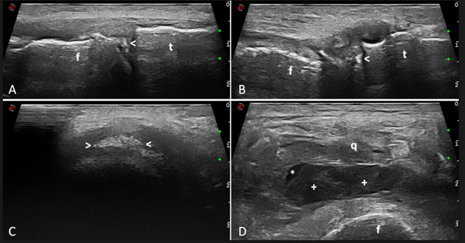
Figure 1: Ultrasound evidence of knee chondrocalcinosis. A-B. Longitudinal medial and lateral scans showing meniscal calcifications appearing as hyperechoic spots not generating acoustic shadowing (arrowheads). C. Axial scan confirming calcifications of the medial meniscus (arrowheads). D. Transverse suprapatellar view showing enlargement of the suprapatellar pouch due to hypoechoic synovial hypertrophy (crosses) and a small amount of anechoic fluid (asterisk). f=femur; q=quadriceps tendon; t=tibia.
Nevertheless, a neither haematic nor purulent synovial fluid was aspirated under ultrasound guidance from the right suprapatellar pouch using an 18-gauge needle and 40 mg of triamcinolone acetonide were injected. Synovial fluid analysis confirmed the diagnosis of CPPD disease and synovial fluid cultures remained negative. An x-ray of the right knee was asked, with the main aim to assess the degree of knee osteoarthritis, and colchicine treatment 1 mg a day was started [1]. At follow-up visit after one week, the patient did not report any intolerance reactions to colchicine (abdominal pain, diarrhea). Clinical and imaging findings led to the request for an intervention by the physiatrist and the orthopaedic surgeon. In fact, while the right knee completely recovered (patient was not complaining of knee pain and no signs of joint inflammation were detectable), two physical findings became evident: first the patient lying on the bed could not lift-up the lower limb and second a lump on the internal aspect and distal part of the thigh was noted which resulted painless and with a tense elastic consistency on palpation (Figure 2)

Figure 2: Right knee. Hypotrophy of the quadriceps muscle (arrowheads) and a lump on the internal distal aspect of the thigh (arrow).
Physiatry
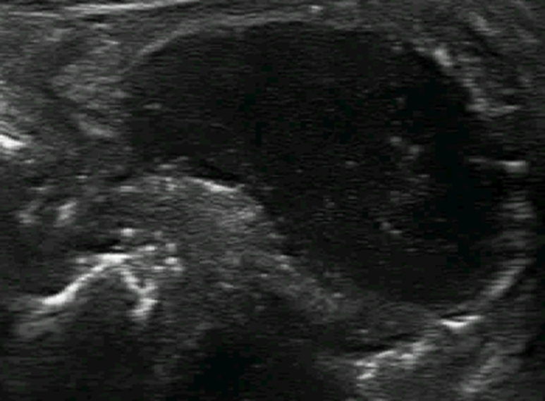
Main findings on physical examination included: diffuse muscular hypotrophy, especially of the right thigh, with right lower limb slightly extra-rotated; active range of motion of the hips extremely limited, especially on the right side; passive right hip mobilization inducing intense antero-lateral thigh pain. The patient exhibited a clear case of secondary sarcopenia, characterized by reduced muscle strength as well as a decrease in both the quantity and quality of muscle mass recorded on both clinical and US examinations. Moreover, in the lower medial third of the thigh, a cold, soft ad mobile mass was noted which showed a tense and elastic consistency on palpation. Thigh pain of the patient did not fit the radicular pattern and did not spread under the knee. No dysesthetic symptoms were reported. The right knee showed no signs of synovitis. Patient lying on the bed could not lift-up the right lower limb and could not stand-up and walk independently. US examination of the thigh revealed a wide quadriceps muscle lesion especially involving vastus medialis, (Figure 3 A-B and Supplementary videos 1 and 2).

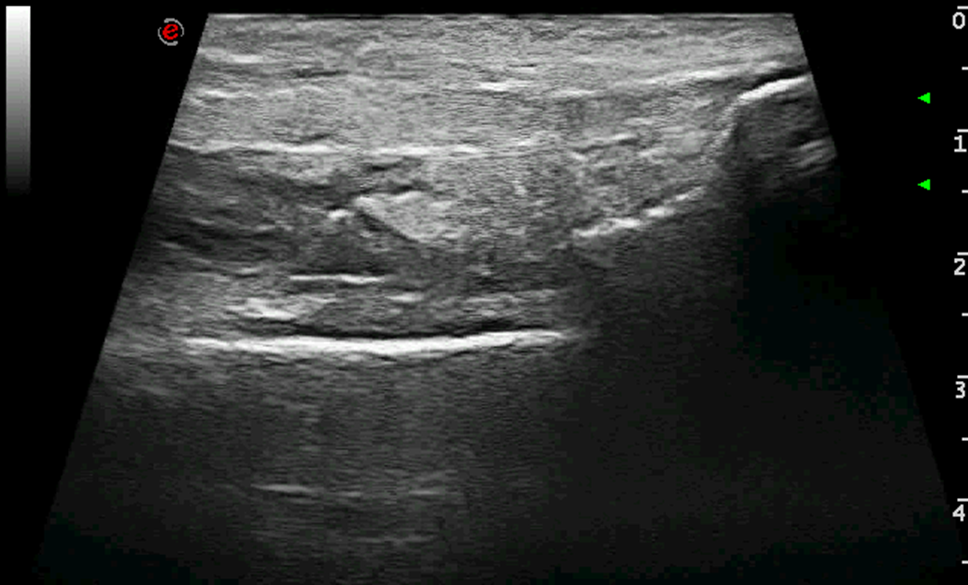
Figure 3: Ultrasound images acquired at baseline (T0) and showing quadriceps muscle rupture. A-B. Longitudinal and transverse scans of the quadriceps muscle showing anechoic fluid collection within the muscle interrupting the tissue texture (asterisks). C. Same view of image B. Needle placement within the muscle lesion during the aspiration of the collection ad injection of 5 mg/2 ml of a solution of hydrolyzed bovine collagen-derived low molecular weight peptides. The arrowhead indicates the tip of the needle. f=femur; q=quadriceps muscle.
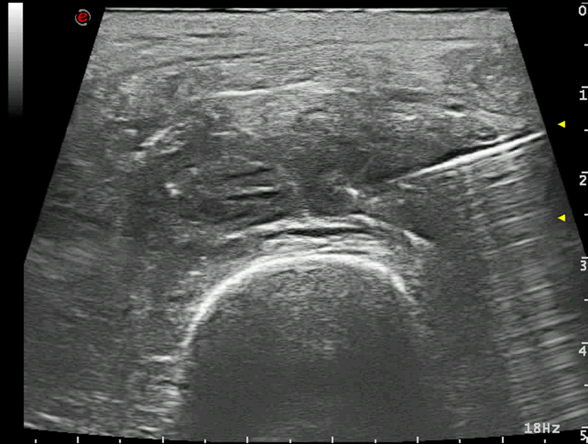
(Main diameters of the lesion: 4.7 x 1.8 x 1.5 cm) and a periosteal cyst (Figure 4 and Supplementary videos 3 and 4).
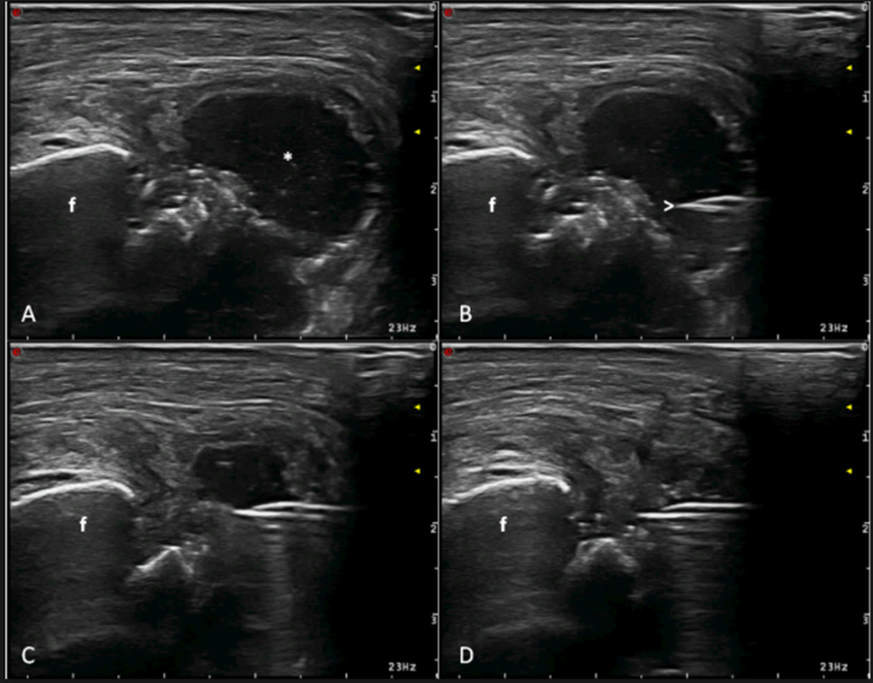
Figure 4: Ultrasound images showing the periosteal abscess (A) and different steps of its ultrasound guided aspiration (B-D). A. Transverse medial scan of the internal distal aspect of the femur showing a hypoechoic round fluid collection (asterisk). B. Tip of the needle placement within the abscess (arrowhead). C. Ultrasound guidance allows for the visualization of the tip of the needle that during the aspiration is maintained in the right position. D. Complete aspiration of the abscess. f=femur.
(Main diameters of the lesion: 3 x 2 x 1.5 cm). The muscle injury was characterized by two large, round-shaped, communicating areas with irregular margins and hypo-anechoic content consistent with a fluid collection. Moreover, X-ray of the knee confirmed CPPD disease and showed bony cortex resorption adjacent to the proximal screw (Figure 5).
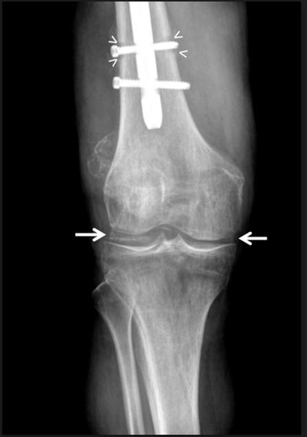
Figure 5: Conventional radiography anteroposterior view of the knee showing meniscal chondrocalcinosis (arrows) and bony cortex resorption adjacent to the proximal screw (arrowheads).
US-guided aspirations of both the fluid collections identified by US were performed. From the quadriceps muscle lesion, the procedure allowed the collection of 8 ml of slightly haematic fluid (Figure 6).
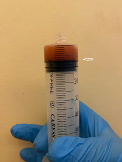
Figure 6: 8 ml of slightly haematic fluid collected from quadriceps muscle lesion (arrow).
while from the periosteal cyst 6 ml of frankly purulent material was aspired, characteristic for abscess and tissue necrosis (Figure 4 B-C-D).
Patient was immediately started with empiric antibiotic therapy, and after five days, following the identification of Serratia marcescens on cultural examination of periosteal cyst, the antibiotic treatment was modified according to the antibiogram: meropenem 1 gr tid and ciprofloxacin 500 mg bid intravenously administered. At home antibiotic therapy continued with cefditorelpivoxil 400 mg sid + trimethoprim-sulfamethoxazole 160/800 mg bid orally. In order to increase muscle mass and strength, patient also took essential amino acid at home [6,7]. The cultures of the haematic fluid aspired from the quadriceps muscle lesion remained negative. Moreover, the patient underwent a personalized rehabilitation treatment under the supervision and assistance of a dedicated physiotherapist. He continued the rehabilitation program independently at home, performing lower limb mobilization exercises, isometric muscle strengthening, and ambulation with partial weight-bearing using a forearm walker.
Intramuscular haematoma US-guided injection with low molecular weight hydrolized bovine collagen solution.
At baseline (T0), after obtaining the patient's informed consent and according to the indications provided by the research and development and drug safety department of HLC Srl, we proceeded with the US-guided aspiration of the quadriceps muscle haematoma and US-guided injection of 5 mg/2 ml of a solution of hydrolyzed bovine collagen-derived low molecular weight peptides (LWPs), (PEPTYS 52©, Horizon Lab Company Srl, San Marino Republic), was performed (Figure 3C and Supplementary video 5). After the procedure, patient underwent a compressive medication and performed a rehabilitation program consisting of hip and knee joint mobilization, isometric quadriceps strengthening and assisted gait rehabilitation program.
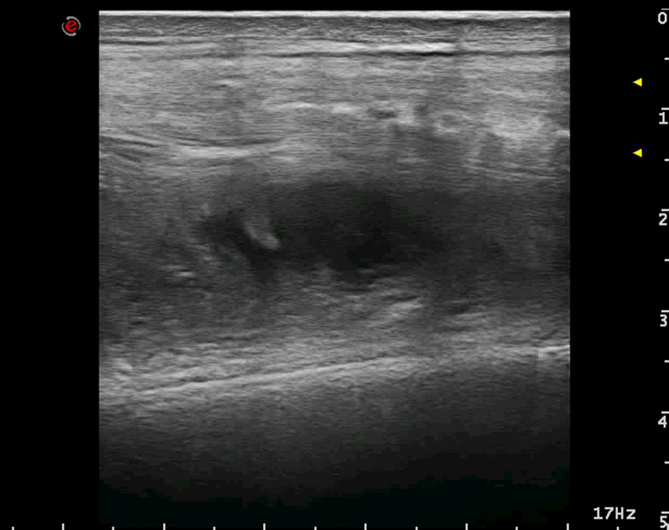
Two weeks after the baseline (T1), patient showed an increase in quadriceps strength and pain during hip flexion reduced; however, he could not stand up nor walking. At T1, US examination showed (Figure 7 A-B) a significant reduction of the muscular lesion and a second injection with LWPs was performed under US guidance.
At week seven from T0, one month after the second injection with LWPs (T2), muscular strength and active range of movement of the right hip increased, pain during hip flexion nearly disappeared and patient was able to stand-up on his own and move independently from bed to wheelchair. At T2, US examination showed further progress in the healing process in terms of volume reduction and echogenicity increase of the muscle lesion due to scar formation (Figure 7 C-D).
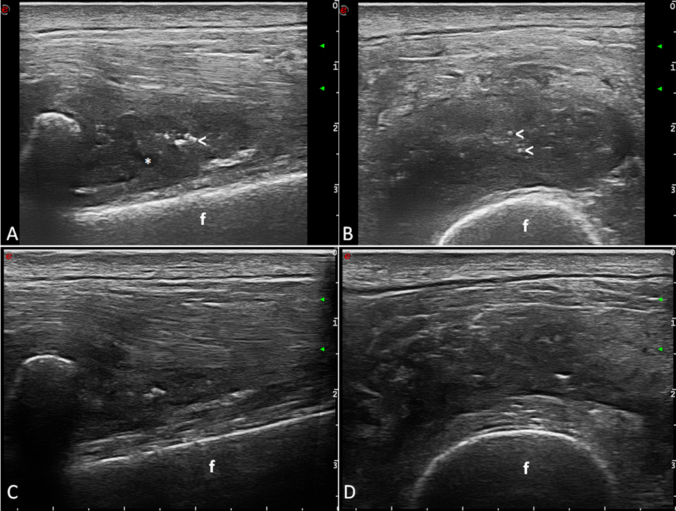
Figure 7: Representative ultrasound images of quadriceps muscle lesion at follow-up visit T1 (A-B) and T2 (C-D), respectively two and six weeks after the first injection of 5 mg/2 ml of a solution of hydrolyzed bovine collagen-derived low molecular weight peptides. A-B. At T1, the muscle lesion appeared more echogenic, some hyperechoic spots (arrowheads) were detected, and anechoic fluid collection was nearly disappeared (asterisk). C-D. At T2, further progress of the healing process could be detected by ultrasound: the volume of the muscle lesion reduced and no fluid collection was detected within the quadriceps muscle.
Figure 7: Representative ultrasound images of quadriceps muscle lesion at follow-up visit T1 (A-B) and T2 (C-D), respectively two and six weeks after the first injection of 5 mg/2 ml of a solution of hydrolyzed bovine collagen-derived low molecular weight peptides. A-B. At T1, the muscle lesion appeared more echogenic, some hyperechoic spots (arrowheads) were detected, and anechoic fluid collection was nearly disappeared (asterisk). C-D. At T2, further progress of the healing process could be detected by ultrasound: the volume of the muscle lesion reduced and no fluid collection was detected within the quadriceps muscle.
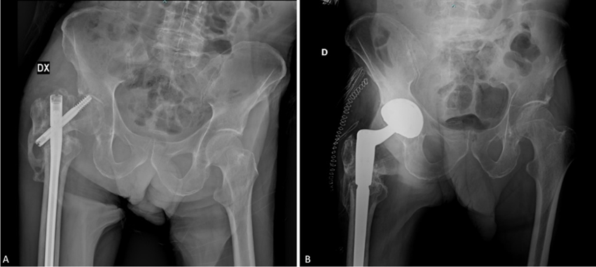
Figure 8: X-ray imaging showing femoral gamma nail (A) and total hip arthroplasty (B).
Description of the surgical intervention: the patient was positioned in left lateral decubitus. A sterile field was prepared with iodine-impregnated adhesive drape. A posterior-lateral incision was made following the Gibson-Moore approach. Fasciotomy was performed, followed by dissection of the extra-rotators. Upon reaching the articular plane, the presence of purulent material was observed, which was removed and sent for culture examination. A pseudarthrosis was identified. The metal implant was removed, along with the gamma nail, using appropriate instruments. The fracture site was regularized, and the acetabulum was prepared using progressively larger reamers up to size 54. The femur was prepared. After adequate debridement, an antibiotic spacer with gentamicin and vancomycin was cemented at the metaphyseal level. Reduction and final stability were tested. A suction drainage system was placed subfascially, followed by layered closure and sterile dressing.
After 6 months, the spacer was removed, and a hip arthroplasty (hip prosthesis) was placed (Figure 8B and 9). Postoperative swab tests were negative. The patient is ambulating with the assistance of a walker.

Figure 9: X-ray imaging showing total hip arthroplasty.
Surgical intervention summary: the procedure was performed with the patient in left lateral decubitus, following a standard posterior-lateral (Gibson-Moore) approach through the previous scar. After removal of the antibiotic spacer and sampling of periprosthetic tissue for cultures, a total hip revision arthroplasty was performed with implantation of a cementless acetabular cup and femoral stem. Stability was confirmed intraoperatively, and the wound was closed in layers with drainage and sterile dressing.
Of note, lavage was performed thoroughly using saline solution. Antibiotic-loaded cement was applied, and proper debridement of the infected tissues was completed. Stability was confirmed after the first test. The postoperative course was uneventful, the patient underwent adequate perioperative care and rehabilitation treatment according to the established protocol [8].
Comprehensive clinical examination and merging clinical with imaging findings were the keys for the accurate diagnosis of this severe and disabling pain syndrome affecting the right lower limb.
In fact, obvious clinical signs of acute knee monoarthritis together with US findings confirming CPPD disease could lead to a hasty conclusion of the diagnostic process neglecting other important clinical signs which could not be explained with knee synovitis [2,3]. As a matter of fact, pain induced by passive right hip mobilization and painless lump on the internal distal aspect of the thigh were due to other pathologic conditions and imaging findings helped in making the correct diagnosis.
In this case, US examination accelerated the diagnostic process by simultaneously identifying three distinct pathological conditions in the right lower limb: knee chondrocalcinosis, quadriceps muscle lesion, and peri-femoral abscess. Additionally, US guidance was crucial for therapeutic interventions, allowing accurate aspiration of fluids and injection of LWPs precisely into the injured muscle area. Conventional radiography later supported US findings, confirming CPPD disease and highlighting radiolucency in the femoral cortex suggestive of infection around the screw.
Moreover, the treatment of a large muscle injury, particularly in a elderly patient with multiple comorbidities and limited recovery capacity, represents a challenge for the physicians.
In recent years, the literature has highlighted the important role of LWPs in soft tissues regeneration such as tendons and cartilage. LWPs can be administered orally or directly injected into the target area [9,10,11]. This innovative therapy has demonstrated a potential role in enhancing muscle performance and in the prevention of soft tissue injuries [10].
However, the efficacy of collagen peptides administered for muscle injury has not yet been described in the literature.
Our therapeutic approach was based on the evidence that the role of the extracellular matrix (ECM) in muscle repair mechanisms is widely recognized [12] and the stimulation of ECM synthesis is probably caused by specific Hyp-Pro-Gly-containing peptides with a molecular size <10>
This case highlighted the healing of the muscle injury both anatomically, through US examination, and functionally, through clinical assessment. Moreover, thanks to the rehabilitation program, the patient managed to increase segmental muscle strength and endurance, to recover quadriceps strength and to restore the functionality of the right lower limb. The rehabilitation treatment also focused on addressing generalized fatigue and counteracting sarcopenia.
In patients presenting with multiple coexisting musculoskeletal conditions—such as peri-implant infection, muscle injury, and chondrocalcinosis—a multidisciplinary and holistic approach is essential. These complex clinical scenarios require not only the integration of expertise from rheumatologists, physiatrists, and orthopaedic surgeons, but also continuous clinical monitoring to promptly identify complications and adjust the therapeutic strategy accordingly. A comprehensive, patient-centered management plan is fundamental to address the interplay of comorbidities and to ensure optimal functional recovery.
We would like to emphasize that the clinical outcome achieved in our case should be considered within the context of a multidisciplinary approach involving a rheumatologist, physiatrist, and orthopedic specialist.
Our work mainly aimed to highlight, on one hand, the significant diagnostic and therapeutic value of value of ultrasound across various medical specialties, and on the other, the importance of teamwork. It is widely recognized that a multidisciplinary approach allows for better management of clinical cases that are challenging to address in routine clinical practice.
We present a case in which the contribution of three different musculoskeletal specialists was essential to resolving the clinical issues encountered.
Although a consensus on the routine use of musculoskeletal ultrasound among specialists in locomotor system disorders—such as physiatrists, orthopaedic surgeons, sports medicine physicians, and rheumatologists—is yet to be fully established, we believe that every effort should be made to promote the widespread adoption of this diagnostic and therapeutic tool. Its broad clinical utility, non-invasiveness, and relatively low cost make it an invaluable resource in daily practice.
This case report supports the integration of musculoskeletal ultrasound and injectable collagen peptides as effective tools in the treatment of soft tissue lesions. The clinical experience described confirms their therapeutic value in enhancing diagnostic precision and promoting tissue recovery. Given their non-invasiveness, safety profile, and growing evidence in the literature, these approaches should be more widely adopted in routine practice. Nevertheless, further studies are needed to establish standardized protocols and optimize their use across different clinical settings.
Supplementary videos legends.
Supplementary Video 1.
Ultrasound imaging of the thigh. Video acquired first placing the probe on the longitudinal anterior scan of the suprapatellar pouch of the knee and then moving the probe proximally towards the hip. The video shows the suprapatellar pouch enlargement, the quadriceps muscle marked hypotrophy and finally the vastus lateralis muscle lesion.
Supplementary Video 2.
The dynamic compression with the ultrasound probe on the thigh showed the displacement of the anechoic area and the movement of the torn muscle flaps. Such ultrasound imaging evidence during real-time examination provides visual confirmation of fluid collection within the quadriceps muscle lesion.
Supplementary Video 3.
The dynamic compression with the ultrasound probe on the lump on the internal distal aspect of the thigh showed the displacement of the hypoechoic material, providing evidence of a fluid collection in communication with the internal part of the bone.

Supplementary Video 4.
Video showing the ultrasound guided aspiration of the periosteal abscess.
Supplementary Video 5.
Video showing the ultrasound guided injection of 5 mg/2 ml of a solution of hydrolyzed bovine collagen-derived low molecular weight peptides within the muscle lesion.
Clearly Auctoresonline and particularly Psychology and Mental Health Care Journal is dedicated to improving health care services for individuals and populations. The editorial boards' ability to efficiently recognize and share the global importance of health literacy with a variety of stakeholders. Auctoresonline publishing platform can be used to facilitate of optimal client-based services and should be added to health care professionals' repertoire of evidence-based health care resources.

Journal of Clinical Cardiology and Cardiovascular Intervention The submission and review process was adequate. However I think that the publication total value should have been enlightened in early fases. Thank you for all.

Journal of Women Health Care and Issues By the present mail, I want to say thank to you and tour colleagues for facilitating my published article. Specially thank you for the peer review process, support from the editorial office. I appreciate positively the quality of your journal.

Journal of Clinical Research and Reports I would be very delighted to submit my testimonial regarding the reviewer board and the editorial office. The reviewer board were accurate and helpful regarding any modifications for my manuscript. And the editorial office were very helpful and supportive in contacting and monitoring with any update and offering help. It was my pleasure to contribute with your promising Journal and I am looking forward for more collaboration.

We would like to thank the Journal of Thoracic Disease and Cardiothoracic Surgery because of the services they provided us for our articles. The peer-review process was done in a very excellent time manner, and the opinions of the reviewers helped us to improve our manuscript further. The editorial office had an outstanding correspondence with us and guided us in many ways. During a hard time of the pandemic that is affecting every one of us tremendously, the editorial office helped us make everything easier for publishing scientific work. Hope for a more scientific relationship with your Journal.

The peer-review process which consisted high quality queries on the paper. I did answer six reviewers’ questions and comments before the paper was accepted. The support from the editorial office is excellent.

Journal of Neuroscience and Neurological Surgery. I had the experience of publishing a research article recently. The whole process was simple from submission to publication. The reviewers made specific and valuable recommendations and corrections that improved the quality of my publication. I strongly recommend this Journal.

Dr. Katarzyna Byczkowska My testimonial covering: "The peer review process is quick and effective. The support from the editorial office is very professional and friendly. Quality of the Clinical Cardiology and Cardiovascular Interventions is scientific and publishes ground-breaking research on cardiology that is useful for other professionals in the field.

Thank you most sincerely, with regard to the support you have given in relation to the reviewing process and the processing of my article entitled "Large Cell Neuroendocrine Carcinoma of The Prostate Gland: A Review and Update" for publication in your esteemed Journal, Journal of Cancer Research and Cellular Therapeutics". The editorial team has been very supportive.

Testimony of Journal of Clinical Otorhinolaryngology: work with your Reviews has been a educational and constructive experience. The editorial office were very helpful and supportive. It was a pleasure to contribute to your Journal.

Dr. Bernard Terkimbi Utoo, I am happy to publish my scientific work in Journal of Women Health Care and Issues (JWHCI). The manuscript submission was seamless and peer review process was top notch. I was amazed that 4 reviewers worked on the manuscript which made it a highly technical, standard and excellent quality paper. I appreciate the format and consideration for the APC as well as the speed of publication. It is my pleasure to continue with this scientific relationship with the esteem JWHCI.

This is an acknowledgment for peer reviewers, editorial board of Journal of Clinical Research and Reports. They show a lot of consideration for us as publishers for our research article “Evaluation of the different factors associated with side effects of COVID-19 vaccination on medical students, Mutah university, Al-Karak, Jordan”, in a very professional and easy way. This journal is one of outstanding medical journal.

Dear Hao Jiang, to Journal of Nutrition and Food Processing We greatly appreciate the efficient, professional and rapid processing of our paper by your team. If there is anything else we should do, please do not hesitate to let us know. On behalf of my co-authors, we would like to express our great appreciation to editor and reviewers.

As an author who has recently published in the journal "Brain and Neurological Disorders". I am delighted to provide a testimonial on the peer review process, editorial office support, and the overall quality of the journal. The peer review process at Brain and Neurological Disorders is rigorous and meticulous, ensuring that only high-quality, evidence-based research is published. The reviewers are experts in their fields, and their comments and suggestions were constructive and helped improve the quality of my manuscript. The review process was timely and efficient, with clear communication from the editorial office at each stage. The support from the editorial office was exceptional throughout the entire process. The editorial staff was responsive, professional, and always willing to help. They provided valuable guidance on formatting, structure, and ethical considerations, making the submission process seamless. Moreover, they kept me informed about the status of my manuscript and provided timely updates, which made the process less stressful. The journal Brain and Neurological Disorders is of the highest quality, with a strong focus on publishing cutting-edge research in the field of neurology. The articles published in this journal are well-researched, rigorously peer-reviewed, and written by experts in the field. The journal maintains high standards, ensuring that readers are provided with the most up-to-date and reliable information on brain and neurological disorders. In conclusion, I had a wonderful experience publishing in Brain and Neurological Disorders. The peer review process was thorough, the editorial office provided exceptional support, and the journal's quality is second to none. I would highly recommend this journal to any researcher working in the field of neurology and brain disorders.
Dear Agrippa Hilda, Journal of Neuroscience and Neurological Surgery, Editorial Coordinator, I trust this message finds you well. I want to extend my appreciation for considering my article for publication in your esteemed journal. I am pleased to provide a testimonial regarding the peer review process and the support received from your editorial office. The peer review process for my paper was carried out in a highly professional and thorough manner. The feedback and comments provided by the authors were constructive and very useful in improving the quality of the manuscript. This rigorous assessment process undoubtedly contributes to the high standards maintained by your journal.

International Journal of Clinical Case Reports and Reviews. I strongly recommend to consider submitting your work to this high-quality journal. The support and availability of the Editorial staff is outstanding and the review process was both efficient and rigorous.

Thank you very much for publishing my Research Article titled “Comparing Treatment Outcome Of Allergic Rhinitis Patients After Using Fluticasone Nasal Spray And Nasal Douching" in the Journal of Clinical Otorhinolaryngology. As Medical Professionals we are immensely benefited from study of various informative Articles and Papers published in this high quality Journal. I look forward to enriching my knowledge by regular study of the Journal and contribute my future work in the field of ENT through the Journal for use by the medical fraternity. The support from the Editorial office was excellent and very prompt. I also welcome the comments received from the readers of my Research Article.

Dear Erica Kelsey, Editorial Coordinator of Cancer Research and Cellular Therapeutics Our team is very satisfied with the processing of our paper by your journal. That was fast, efficient, rigorous, but without unnecessary complications. We appreciated the very short time between the submission of the paper and its publication on line on your site.

I am very glad to say that the peer review process is very successful and fast and support from the Editorial Office. Therefore, I would like to continue our scientific relationship for a long time. And I especially thank you for your kindly attention towards my article. Have a good day!

"We recently published an article entitled “Influence of beta-Cyclodextrins upon the Degradation of Carbofuran Derivatives under Alkaline Conditions" in the Journal of “Pesticides and Biofertilizers” to show that the cyclodextrins protect the carbamates increasing their half-life time in the presence of basic conditions This will be very helpful to understand carbofuran behaviour in the analytical, agro-environmental and food areas. We greatly appreciated the interaction with the editor and the editorial team; we were particularly well accompanied during the course of the revision process, since all various steps towards publication were short and without delay".

I would like to express my gratitude towards you process of article review and submission. I found this to be very fair and expedient. Your follow up has been excellent. I have many publications in national and international journal and your process has been one of the best so far. Keep up the great work.

We are grateful for this opportunity to provide a glowing recommendation to the Journal of Psychiatry and Psychotherapy. We found that the editorial team were very supportive, helpful, kept us abreast of timelines and over all very professional in nature. The peer review process was rigorous, efficient and constructive that really enhanced our article submission. The experience with this journal remains one of our best ever and we look forward to providing future submissions in the near future.

I am very pleased to serve as EBM of the journal, I hope many years of my experience in stem cells can help the journal from one way or another. As we know, stem cells hold great potential for regenerative medicine, which are mostly used to promote the repair response of diseased, dysfunctional or injured tissue using stem cells or their derivatives. I think Stem Cell Research and Therapeutics International is a great platform to publish and share the understanding towards the biology and translational or clinical application of stem cells.

I would like to give my testimony in the support I have got by the peer review process and to support the editorial office where they were of asset to support young author like me to be encouraged to publish their work in your respected journal and globalize and share knowledge across the globe. I really give my great gratitude to your journal and the peer review including the editorial office.

I am delighted to publish our manuscript entitled "A Perspective on Cocaine Induced Stroke - Its Mechanisms and Management" in the Journal of Neuroscience and Neurological Surgery. The peer review process, support from the editorial office, and quality of the journal are excellent. The manuscripts published are of high quality and of excellent scientific value. I recommend this journal very much to colleagues.

Dr.Tania Muñoz, My experience as researcher and author of a review article in The Journal Clinical Cardiology and Interventions has been very enriching and stimulating. The editorial team is excellent, performs its work with absolute responsibility and delivery. They are proactive, dynamic and receptive to all proposals. Supporting at all times the vast universe of authors who choose them as an option for publication. The team of review specialists, members of the editorial board, are brilliant professionals, with remarkable performance in medical research and scientific methodology. Together they form a frontline team that consolidates the JCCI as a magnificent option for the publication and review of high-level medical articles and broad collective interest. I am honored to be able to share my review article and open to receive all your comments.

“The peer review process of JPMHC is quick and effective. Authors are benefited by good and professional reviewers with huge experience in the field of psychology and mental health. The support from the editorial office is very professional. People to contact to are friendly and happy to help and assist any query authors might have. Quality of the Journal is scientific and publishes ground-breaking research on mental health that is useful for other professionals in the field”.

Dear editorial department: On behalf of our team, I hereby certify the reliability and superiority of the International Journal of Clinical Case Reports and Reviews in the peer review process, editorial support, and journal quality. Firstly, the peer review process of the International Journal of Clinical Case Reports and Reviews is rigorous, fair, transparent, fast, and of high quality. The editorial department invites experts from relevant fields as anonymous reviewers to review all submitted manuscripts. These experts have rich academic backgrounds and experience, and can accurately evaluate the academic quality, originality, and suitability of manuscripts. The editorial department is committed to ensuring the rigor of the peer review process, while also making every effort to ensure a fast review cycle to meet the needs of authors and the academic community. Secondly, the editorial team of the International Journal of Clinical Case Reports and Reviews is composed of a group of senior scholars and professionals with rich experience and professional knowledge in related fields. The editorial department is committed to assisting authors in improving their manuscripts, ensuring their academic accuracy, clarity, and completeness. Editors actively collaborate with authors, providing useful suggestions and feedback to promote the improvement and development of the manuscript. We believe that the support of the editorial department is one of the key factors in ensuring the quality of the journal. Finally, the International Journal of Clinical Case Reports and Reviews is renowned for its high- quality articles and strict academic standards. The editorial department is committed to publishing innovative and academically valuable research results to promote the development and progress of related fields. The International Journal of Clinical Case Reports and Reviews is reasonably priced and ensures excellent service and quality ratio, allowing authors to obtain high-level academic publishing opportunities in an affordable manner. I hereby solemnly declare that the International Journal of Clinical Case Reports and Reviews has a high level of credibility and superiority in terms of peer review process, editorial support, reasonable fees, and journal quality. Sincerely, Rui Tao.

Clinical Cardiology and Cardiovascular Interventions I testity the covering of the peer review process, support from the editorial office, and quality of the journal.

Clinical Cardiology and Cardiovascular Interventions, we deeply appreciate the interest shown in our work and its publication. It has been a true pleasure to collaborate with you. The peer review process, as well as the support provided by the editorial office, have been exceptional, and the quality of the journal is very high, which was a determining factor in our decision to publish with you.

The peer reviewers process is quick and effective, the supports from editorial office is excellent, the quality of journal is high. I would like to collabroate with Internatioanl journal of Clinical Case Reports and Reviews journal clinically in the future time.

Clinical Cardiology and Cardiovascular Interventions, I would like to express my sincerest gratitude for the trust placed in our team for the publication in your journal. It has been a true pleasure to collaborate with you on this project. I am pleased to inform you that both the peer review process and the attention from the editorial coordination have been excellent. Your team has worked with dedication and professionalism to ensure that your publication meets the highest standards of quality. We are confident that this collaboration will result in mutual success, and we are eager to see the fruits of this shared effort.

Dear Dr. Jessica Magne, Editorial Coordinator 0f Clinical Cardiology and Cardiovascular Interventions, I hope this message finds you well. I want to express my utmost gratitude for your excellent work and for the dedication and speed in the publication process of my article titled "Navigating Innovation: Qualitative Insights on Using Technology for Health Education in Acute Coronary Syndrome Patients." I am very satisfied with the peer review process, the support from the editorial office, and the quality of the journal. I hope we can maintain our scientific relationship in the long term.

Dear Monica Gissare, - Editorial Coordinator of Nutrition and Food Processing. ¨My testimony with you is truly professional, with a positive response regarding the follow-up of the article and its review, you took into account my qualities and the importance of the topic¨.

Dear Dr. Jessica Magne, Editorial Coordinator 0f Clinical Cardiology and Cardiovascular Interventions, The review process for the article “The Handling of Anti-aggregants and Anticoagulants in the Oncologic Heart Patient Submitted to Surgery” was extremely rigorous and detailed. From the initial submission to the final acceptance, the editorial team at the “Journal of Clinical Cardiology and Cardiovascular Interventions” demonstrated a high level of professionalism and dedication. The reviewers provided constructive and detailed feedback, which was essential for improving the quality of our work. Communication was always clear and efficient, ensuring that all our questions were promptly addressed. The quality of the “Journal of Clinical Cardiology and Cardiovascular Interventions” is undeniable. It is a peer-reviewed, open-access publication dedicated exclusively to disseminating high-quality research in the field of clinical cardiology and cardiovascular interventions. The journal's impact factor is currently under evaluation, and it is indexed in reputable databases, which further reinforces its credibility and relevance in the scientific field. I highly recommend this journal to researchers looking for a reputable platform to publish their studies.

Dear Editorial Coordinator of the Journal of Nutrition and Food Processing! "I would like to thank the Journal of Nutrition and Food Processing for including and publishing my article. The peer review process was very quick, movement and precise. The Editorial Board has done an extremely conscientious job with much help, valuable comments and advices. I find the journal very valuable from a professional point of view, thank you very much for allowing me to be part of it and I would like to participate in the future!”

Dealing with The Journal of Neurology and Neurological Surgery was very smooth and comprehensive. The office staff took time to address my needs and the response from editors and the office was prompt and fair. I certainly hope to publish with this journal again.Their professionalism is apparent and more than satisfactory. Susan Weiner

My Testimonial Covering as fellowing: Lin-Show Chin. The peer reviewers process is quick and effective, the supports from editorial office is excellent, the quality of journal is high. I would like to collabroate with Internatioanl journal of Clinical Case Reports and Reviews.

My experience publishing in Psychology and Mental Health Care was exceptional. The peer review process was rigorous and constructive, with reviewers providing valuable insights that helped enhance the quality of our work. The editorial team was highly supportive and responsive, making the submission process smooth and efficient. The journal's commitment to high standards and academic rigor makes it a respected platform for quality research. I am grateful for the opportunity to publish in such a reputable journal.

My experience publishing in International Journal of Clinical Case Reports and Reviews was exceptional. I Come forth to Provide a Testimonial Covering the Peer Review Process and the editorial office for the Professional and Impartial Evaluation of the Manuscript.

I would like to offer my testimony in the support. I have received through the peer review process and support the editorial office where they are to support young authors like me, encourage them to publish their work in your esteemed journals, and globalize and share knowledge globally. I really appreciate your journal, peer review, and editorial office.

Dear Agrippa Hilda- Editorial Coordinator of Journal of Neuroscience and Neurological Surgery, "The peer review process was very quick and of high quality, which can also be seen in the articles in the journal. The collaboration with the editorial office was very good."

I would like to express my sincere gratitude for the support and efficiency provided by the editorial office throughout the publication process of my article, “Delayed Vulvar Metastases from Rectal Carcinoma: A Case Report.” I greatly appreciate the assistance and guidance I received from your team, which made the entire process smooth and efficient. The peer review process was thorough and constructive, contributing to the overall quality of the final article. I am very grateful for the high level of professionalism and commitment shown by the editorial staff, and I look forward to maintaining a long-term collaboration with the International Journal of Clinical Case Reports and Reviews.

To Dear Erin Aust, I would like to express my heartfelt appreciation for the opportunity to have my work published in this esteemed journal. The entire publication process was smooth and well-organized, and I am extremely satisfied with the final result. The Editorial Team demonstrated the utmost professionalism, providing prompt and insightful feedback throughout the review process. Their clear communication and constructive suggestions were invaluable in enhancing my manuscript, and their meticulous attention to detail and dedication to quality are truly commendable. Additionally, the support from the Editorial Office was exceptional. From the initial submission to the final publication, I was guided through every step of the process with great care and professionalism. The team's responsiveness and assistance made the entire experience both easy and stress-free. I am also deeply impressed by the quality and reputation of the journal. It is an honor to have my research featured in such a respected publication, and I am confident that it will make a meaningful contribution to the field.

"I am grateful for the opportunity of contributing to [International Journal of Clinical Case Reports and Reviews] and for the rigorous review process that enhances the quality of research published in your esteemed journal. I sincerely appreciate the time and effort of your team who have dedicatedly helped me in improvising changes and modifying my manuscript. The insightful comments and constructive feedback provided have been invaluable in refining and strengthening my work".

I thank the ‘Journal of Clinical Research and Reports’ for accepting this article for publication. This is a rigorously peer reviewed journal which is on all major global scientific data bases. I note the review process was prompt, thorough and professionally critical. It gave us an insight into a number of important scientific/statistical issues. The review prompted us to review the relevant literature again and look at the limitations of the study. The peer reviewers were open, clear in the instructions and the editorial team was very prompt in their communication. This journal certainly publishes quality research articles. I would recommend the journal for any future publications.

Dear Jessica Magne, with gratitude for the joint work. Fast process of receiving and processing the submitted scientific materials in “Clinical Cardiology and Cardiovascular Interventions”. High level of competence of the editors with clear and correct recommendations and ideas for enriching the article.

We found the peer review process quick and positive in its input. The support from the editorial officer has been very agile, always with the intention of improving the article and taking into account our subsequent corrections.

My article, titled 'No Way Out of the Smartphone Epidemic Without Considering the Insights of Brain Research,' has been republished in the International Journal of Clinical Case Reports and Reviews. The review process was seamless and professional, with the editors being both friendly and supportive. I am deeply grateful for their efforts.

To Dear Erin Aust – Editorial Coordinator of Journal of General Medicine and Clinical Practice! I declare that I am absolutely satisfied with your work carried out with great competence in following the manuscript during the various stages from its receipt, during the revision process to the final acceptance for publication. Thank Prof. Elvira Farina

Dear Jessica, and the super professional team of the ‘Clinical Cardiology and Cardiovascular Interventions’ I am sincerely grateful to the coordinated work of the journal team for the no problem with the submission of my manuscript: “Cardiometabolic Disorders in A Pregnant Woman with Severe Preeclampsia on the Background of Morbid Obesity (Case Report).” The review process by 5 experts was fast, and the comments were professional, which made it more specific and academic, and the process of publication and presentation of the article was excellent. I recommend that my colleagues publish articles in this journal, and I am interested in further scientific cooperation. Sincerely and best wishes, Dr. Oleg Golyanovskiy.
