AUCTORES
Globalize your Research
Research Article | DOI: https://doi.org/10.31579/2688-7517/197
Faculdade Ciências Médicas de Minas Gerais
*Corresponding Author: : Thiago Henrique Caldeira de Oliveira, Faculdade Ciências Médicas de Minas Gerais, Alameda Ezequiel Dias, 275 - Centro, Belo Horizonte - MG, 30130-110.
Citation: Caldeira De Oliveira TH, Neves Gonçalves GK, (2025), Effect of Ovariectomy and High-fat Diet on the Expression of Estrogen Receptors and Adipose Tissue Metabolism in Wistar Rats, J. Pharmaceutics and Pharmacology Research, 8(1); DOI:10.31579/2688-7517/197
Copyright: © 2025, Thiago Henrique Caldeira de Oliveira. This is an open access article distributed under the Creative Commons Attribution License, which permits unrestricted use, distribution, and reproduction in any medium, provided the original work is properly cited.
Received: 29 May 2024 | Accepted: 01 June 2024 | Published: 13 January 2025
Keywords: adipose tissue; post-menopause; estrogen
Background: This study examines the rising prevalence of obesity in postmenopausal women and its link to hypertension and endothelial dysfunction. Visceral fat poses the highest risk for cardiovascular disease and type 2 diabetes. Estrogen regulates adipose tissue, and its absence post-menopause is linked to metabolic issues.
Aim: The study aimed to determine lipolytic activity in various adipose tissues of high-fat diet-fed ovariectomized Wistar rats, analyze estrogen receptor expression, and perform histological and morphometric analyses of these tissues.
Materials and Methods: 12-week-old female wistar rats were used. The rats were ovariectomized (O) or sham operated (S). Four weeks later, the animals were divided into groups: ovariectomized with high-fat diet (OF), sham-operated with high-fat diet (SF), ovariectomized with control diet (OC) or sham-operated with control diet as the control group (SC). After 24 weeks of consuming the diets, the rats were sacrificed and the mesenteric, parametrial and retroperitoneal adipose tissue deposits were removed, weighed and immediately frozen. Polymerase chain reaction (PCR) was performed to analyze the expression of estrogen receptors alpha (ER-α) and (ER-β) in adipose tissues, lipolysis assay and histological analysis thereof.
Results: The results demonstrated that both the high-fat diet and ovariectomy resulted in increased body weight and adiposity. There was hypertrophy of adipocytes in certain fat deposits. Estrogen replacement therapy was able to modulate lipolytic activity in different adipose depots, with different responses in relation to estrogen receptors. Estrogen receptor expression varied between fat depots and was influenced by diet and ovariectomy. Mesenteric adipose tissue showed greater sensitivity to estrogen compared with parametrial adipose tissue. Estrogen administration increased lipolytic activity in some fat depots, while reducing it in others.
Conclusions: The expression of estrogen receptors is dependent not only on hormonal status, but also on the location of adipose tissue and diet, which may explain the distinct actions of estrogen both on the metabolism of adipose tissue and on the production of adipokines by them.
The prevalence of obesity has increased in recent decades, particularly among postmenopausal women. Obesity is a recognized risk factor for the development of hypertension, often linked with endothelial dysfunction [1-3]. Postmenopause is associated with the development of central obesity, which increases the risk of hypertension, elevated blood lipid levels, insulin resistance, and other cardiovascular diseases [4]. The distribution of adipose tissue has a significant impact on the risks associated with obesity. While visceral fat, which accumulates around the internal organs in the abdominal cavity, is associated with an increased risk of cardiovascular diseases, type 2 diabetes, and metabolic syndrome, subcutaneous fat, which accumulates just below the skin, has a lesser impact on metabolic health [5]. It is suggested that women in the premenopausal stage are protected from these deleterious effects due to the estrogen hormone produced by the ovaries [6].
Estrogen plays a crucial role in regulating the development and storage of adipose tissue in females. Estrogens encompass a group of compounds, comprising estrone, estradiol, and estriol [7]. Estradiol, in particular, can be subdivided into 17 alpha-estradiol (17a-E2) and 17 beta-estradiol (17b-E2) [8]. Among the four types of estrogen, 17β-estradiol, the most potent, exerts its influence through activation of intracellular estrogen receptors known as ER-α and ER-β [9]. After menopause, women tend to increase their adipose mass, largely due to low levels of 17β-estradiol, which may contribute to the development of various chronic diseases, including hypertension. Moreover, Estrogen deprivation has been associated with an increased risk of obesity and metabolic complications, which can be partially reversed by hormone replacement therapy [10].
Despite the epidemiological evidence, it is crucial to conduct additional studies to elucidate the underlying mechanisms by which estrogen influences adipose tissue and the cardiovascular system. Furthermore, it has recently been observed that adipose tissue deposits manifest distinct responses and secretions, varying according to their location in the body [11-13].
The aim of this study was to determine the lipolytic activity in the parametrial, retroperitoneal and mesenteric adipose tissues of ovariectomized Wistar rats submitted to a high-fat diet. In addition, to analyze the expression of estrogen receptors in different types of adipose tissue in animals, as well as histologically and morphometrically analyzing the retroperitoneal, mesenteric and parametrial adipose tissue deposits of ovariectomized Wistar rats fed a high-fat diet.
2.1 Animals
The study was approved by the “Ethics Committee in Animal Experimentation of the Estadual University of Feira de Santana” and carried out according to the recommendations of the National Institutes of Health Guide for the Care and Use of Laboratory Animals. Female Wistar rats weighting 150–200 g was obtained from ICS-UFBA (Salvador, BA, Brazil). The animals (6 per cage) were maintained under controlled light and temperature conditions (lights on from 07:00 to 19:00 h, 23±3 °C) and had free access to tap water and a standard rat chow (AIN 93; Pragsoluções, Brazil). The animals were ovariectomized or sham-operated using ketamine 50 mg/kg and xylazine 5 mg/kg (im). The animals received a prophylactic antibiotic injection (pentabiotic, Fontoura-Wyeth, 0.4 ml/animal/im) [14]. There were no specific exclusion criteria for this study. All animals that meet the inclusion criteria are eligible to participate
2.2 Diet Protocol
After 1-week acclimation, rats were randomly divided into four groups: ovariectomized with high-fat diet (OF, n=6), sham-operated with high-fat diet (SF, n=6), ovariectomized with control diet (OC, n=6) or sham-operated with control diet as the control group (SC, n=6) [14]. Diets were manufactured by Pragsoluções (São Paulo, SP, Brazil) [15]. Body weight and blood pressure were measured weekly for 24 weeks. Then, estrogen replacement therapy was started. The treated groups received 17β-estradiol (Benzoginoestril; SARSA- Hoechst Marion Roussel, 5mg/mL ampoules), diluted in corn oil at a dose of 5µg/0.1ml per 100g of body weight of benzoginoestril, for fourteen consecutive days. Control animals received 0.1mL of corn oil per 100g of body weight during the same period. The rats were killed by decapitation and the mesenteric, parametrial and retroperitoneal adipose tissue depots were removed, weighed and used for further analysis of gene expression of estrogen receptors as well as histological analysis. Part of the tissues was used immediately for the lipolysis assay [16].
2.3 Adipocyte isolation
Adipocytes were isolated from parametrial, retroperitoneal and mesenteric adipose tissues. After digestion with collagenase, cells were filtered and washed three times with 30mmol/L HEPES buffer (pH 7.4) containing 137mmol/L NaCl, 5mmol/L KCl, 4.2mmol/L NaHCO3, 1.3mmol/L CaCl2, MgCl2 0.5mmol/L, MgSO4 0.5mmol/L, KH2PO4. Then, the cells were resuspended in a proportion of 1g of tissue to 20mL of buffer, so that each mL of medium contained 50mg of cells, which were used for lipolysis studies, measuring cell number and diameter [17].
2.4 Lipolysis assay
Adipocytes were incubated under basal conditions or with isoproterenol (0.1 µM) for 90 minutes. Adipocytes from animals treated with vehicle and 17β-estradiol were used in order to characterize their participation in the process. At the end of the incubation period, an aliquot of the infranatant was collected and the glycerol content analyzed. The glycerol released in the lipolysis incubation medium was quantified by an enzymatic kit (ANALISA, Lagoa Santa, MG) [17, 18].
2.5 Diameter and number of cells
The adipocyte suspension was analyzed in a Newbauer chamber, with the number of cells measured in the four outer quadrants of the chamber, and the cells in the inner quadrant used to analyze cell diameter. To calculate the number of cells, the following formula was used: No cells/mL = (No of cells counted) ÷ (No of quadrants counted) X 104 X dilution factor [19].
2.6 mRNA extraction
Tissues were removed and frozen in liquid nitrogen. Total RNA was extracted from adipose tissues according to the thiocyonate-phenol-chloroform method. Total RNA was extracted using Brazol@ reagent at a proportion of 1ml/100mg of tissue. The RNA concentration was measured in a spectrophotometer at an absorbance of 260nm [16].
2.7 Reverse Transcription
The cDNA was obtained through retro-transcription (RT) from the mRNA, using oligonucleotides complementary to the poly-A tail of the mRNA. For reverse transcription, 4ul of specific buffer (10x), 3uL of dNTP (10mM), 4uL of oligo dt primer and 0.2uL of Moloney Murine Leukemia Virus (MMLV) reverse transcriptase enzyme were added to 1µg of total RNA. The solution was then kept at 40°C for 60 minutes for c-DNA synthesis.
2.8 Polymerase chain reaction (PCR)
PCR was performed to amplify the c-DNA for ER-α and ER-β. To amplify ER-α, the following primers were used: sense primer with sequence TAAGAACCGGAGGAACAGTTG, and antisense primer with sequence TCATGCGGAATCGACTTG (product with 623 base pairs); To amplify ER-β, the following were used: sense primer with sequence GCTCCTCTATGCAGAACCTCAAA, and antisense primer with sequence CAGAAGTGAGCATCCCTCTTTG (product with 159 base pairs). PCR was performed using 20µl of MIX solution containing 2.2µl of specific buffer, 1µl dNTP (4mM), 0.2µl of each primer, 1µl of cDNA, 0.1µl of Taq polymerase and 15.30µl of water. The reaction products were analyzed by electrophoresis on a 1.5% agarose gel. The gel was developed with ethidium bromide (1µg/ml), visualized under ultraviolet light and digitalized. The bands were quantified using the ImageQuant 5 program. β-Actin was amplified as an internal control for each PCR using previously described specific primers. The results refer to three PCRs for each sample [16].
2.9 Morphological and histological analysis
Histological sections of adipose tissue measuring 5 micrometers (µm) thick were evaluated. These sections were subjected to hematoxylin-eosin staining for quantitative tissue analysis. Morphometric measurements were carried out using an interactive computerized image analysis system (Image Pro Plus 4.1 imaging software from Media Cybernetics, Silver Spring, MD).
2.10 Data analysis
Data were expressed as mean ± SEM and analyzed using GraphPad software. Data were compared using one-way ANOVA followed by Newman–Keuls post-hoc test or two-way ANOVA followed by Bonferroni post-test. Correlations were calculated using two-tailed Pearson's correlation coefficients. Data was considered statistically significant at the pb0.05 level.
The initial results demonstrate that both high-fat diet and ovariectomy, alone, resulted in an increase in body weight. Furthermore, the combination of a high-fat diet with ovariectomy resulted in a significant increase in body weight, which was also reflected in the different deposits of adipose tissue (Figure 1A). Ovariectomy resulted in an increase in parametrial fat deposition, while the high-fat diet caused an increase in parametrial and mesenteric fat deposits, compared to control animals. However, neither diet nor ovariectomy influenced the weight of the retroperitoneal fat deposit (Figure 1B).
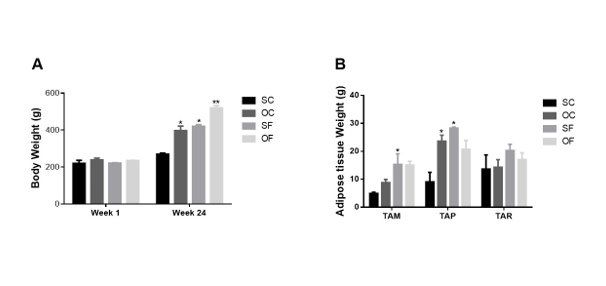
Figure 1: Comparison of weight gain (A) and visceral adipose tissue between animals fed a control and high-fat diet (B) (*p< 0>
The figure 2 shows fat cell morphology after 24 weeks of diet. Control diet animals had uniformly sized adipocytes across all deposits. However, ovariectomized rats on a high-fat diet (OF) exhibited hypertrophic adipocytes in parametrial and mesenteric tissues without increased vasculature, while retroperitoneal tissue showed no significant changes.
The morphological changes in fat distribution induced by a high-fat diet were accompanied by variations in the histological structure of adipose tissue. A high-fat diet caused changes in adipose tissue structure, with the OF group showing increased adipocyte diameter in mesenteric and parametrial tissues, but not in retroperitoneal tissue (Figure 3A). Histological analysis revealed larger adipocytes in mesenteric tissue compared to retroperitoneal and parametrial tissues. Estradiol treatment reduced cell diameter in mesenteric tissue but did not affect parametrial or retroperitoneal adipocytes (Figure 3B).
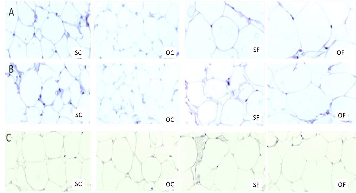
Figure 2: Micrograph of adipose tissue sections from the mesenteric (A), parametrial (B) and retroperitoneal (C) depots of SC, OC, SF and OF rats. Adipose tissue sections from different areas of all deposits were fixed and stained with hematoxylin and eosin. Micrographs were taken at 10x magnitude. Representative sections of each deposit are shown. Micrographs of the parametrial and mesenteric adipose tissue depots of OF rats show hypertrophic adipocytes without evidence of an increase in tissue vasculature. Retroperitoneal adipose tissue does not show any significant changes between groups.
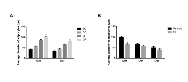
Figure 3: Diameter of adipocytes from the mesenteric (TAM) and parametrial (TAP) adipose tissues of ovariectomized wistar rats fed for 24 weeks with a high-fat diet. * p<0>
A lipolysis assay quantified glycerol release. Isoproterenol, an adrenergic agonist, increased lipolytic activity in all adipose tissues. Retroperitoneal tissue showed higher basal and isoproterenol-stimulated lipolysis. Pretreatment with 17β-estradiol for 14 days reduced lipolysis in retroperitoneal tissue but increased it in parametrial and mesenteric tissues, especially with isoproterenol (Figure 4).
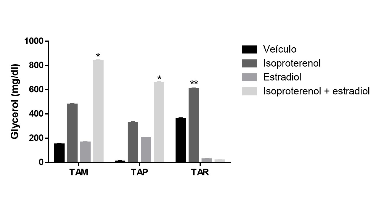
Figure 4: Lipolysis assay of adipocytes isolated from mesenteric (TAM), parametrial (TAP) and retroperitoneal (TAR) adipose tissues of ovariectomized Wistar rats fed a high-fat diet. The results are expressed in mg/dl of glycerol released into the medium under basal conditions or stimulated by isoproterenol. The results refer to the “pool” of adipocytes from five rats treated with vehicle (v) or five rats treated with estradiol (E) for 14 days. *p< 0>
To study the mechanisms involved in the different responses of adipose tissue depots, the gene expression of estrogen receptors was analyzed. Parametrial adipose tissue in control rats showed greater ER-β expression compared to ER-α (Figure 5A), a difference not seen in mesenteric tissue (Figure 5B). After 24 weeks of a high-fat diet, ER-α and ER-β expression in parametrial tissue decreased. Ovariectomy also reduced ER-β expression in this tissue. In contrast, mesenteric tissue showed increased estrogen receptor expression with a high-fat diet, both alone and with ovariectomy.
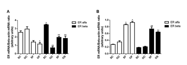
Figure 5: Gene expression of alpha and beta estrogen receptor (ER) in parametrial adipose tissue (TAP) (A) and mesenteric adipose tissue (TAM) (B) of ovariectomized wistar rats fed for 24 weeks with a high-fat diet (OF). * p<0>
Estrogens are considered important regulators of development and lipid deposition in adipose tissue, especially in females. For instance, males typically exhibit a greater propensity for accumulating visceral adiposity, commonly referred to as the "apple shape," heightening the risk of metabolic disorders and cardiometabolic diseases. Conversely, females tend to accrue subcutaneous adipose tissue, providing metabolic protective advantages [20].
To elucidate estrogen's role, estrogen replacement therapy was administered and adipocytes analyzed. Mesenteric adipose tissue, which had the greatest hypertrophy, responded best to therapy, showing a significant reduction in adipocyte diameter after 17β-estradiol treatment.
These data are in line with others found in the literature, which demonstrated that 17β-estradiol decreases cell diameter [21-23]. Furthermore, estradiol replacement therapy in both rodents and humans reverses obesity by reducing visceral fat mass, thereby enhancing metabolic fitness [24, 25]. Increasing evidence from studies conducted in humans and rodents suggests that higher levels of estrogen promote the expansion of subcutaneous adipose tissue while inhibiting the growth of visceral adipose tissue [7]. On the other hand, lower levels of estrogen by menopause or ovariectomy increases the risk of developing obesity, type 2 diabetes and cardiovascular disease [26].
The effects of estrogen on lipid deposition can be attributed to its inhibitory activity on the enzyme lipoprotein lipase (LPL), which regulates lipid uptake by adipocytes. LPL is a crucial enzyme in lipid metabolism. Its main function is to catalyze the hydrolysis of triglycerides present in chylomicrons and very low-density lipoproteins (VLDL), releasing free fatty acids to be absorbed and stored in adipose tissues or used as a source of energy by peripheral tissues [27]. In this regard, when estrogen levels decrease, an increase in LPL activity may occur. Therefore, an increase in LPL activity during menopause may contribute to a greater accumulation of visceral fat and, consequently, increase the risk of developing conditions related to metabolic health [28]. On the other hand, studies demonstrate that estrogen treatment decreases lipid deposition by decreasing LPL activity [29]. Moreover, estrogen markedly decreased the amounts of fat accumulation and LPL mRNA as well as triglyceride accumulation in genetically manipulated 3T3-L1 adipocytes stably expressing the estrogen receptor [30].
Adipose mass reflects both the number and size of the adipocyte, the latter being direct evidence of the stored triacylglycerol content. Thus, the reduction in the mass of parametrial and mesenteric adipose tissue in the OF group treated with estradiol coincides with the reduction in the average diameter of the adipocyte, suggesting that the effect of estradiol in this tissue is mediated, to a large extent, by the effects of this hormone on the adipocyte size. In this regard, studies demonstrate that 17β-Estradiol decreases the intracellular triacylglycerol levels. Moreover, this effect is abolished by ERα antagonist but not ERβ antagonist [31, 32].
The lipolysis assay shows varied tissue responses. Estradiol enhances isoproterenol's effects on mesenteric and parametrial fat, but in parametrial tissue, it acts similarly to isoproterenol alone. In retroperitoneal tissue, estradiol inhibits lipolysis both alone and with isoproterenol. Thus, estradiol increases basal glycerol release from parametrial and mesenteric tissues in rats on a high-fat diet. Similar results were observed in female wistar rats during diestrus, as well as in in situ microdialysis studies performed in ovariectomized females after estrogen administration [9, 33]. These results implicate estrogens as modulators of basal and stimulated lipolytic responses in rat visceral fat in vivo.
It was also observed that adrenergic stimulation by isoproterenol was not influenced differently by hormone replacement in retroperitoneal deposits, corroborating results obtained in women, in whom estradiol replacement therapy did not alter the lipolytic activity stimulated by noradrenaline in subcutaneous adipocytes [34]. These data show that retroperitoneal adipose tissue appears to behave differently from other visceral tissues, resembling subcutaneous adipose tissue. In this way, the different localization sites of adipose tissue in the body seem to perform their functions, including lipolysis and lipogenesis in different ways, as well as responding to external stimuli, such as diet, in different ways. In this regard, other studies corroborate this work, which show that the volume of adipocytes in visceral adipose tissue is greater when compared to those in subcutaneous tissue. Furthermore, adipose tissue depots have different sensitivities to important hormones that regulate adipose tissue metabolism. Subcutaneous adipose tissue appears to be less sensitive to epinephrine and norepinephrine when compared to mesenteric adipose tissue, and may have 50% less lipolysis [35]. Catecholamines are potent activators of lipolysis that act on adrenergic receptors, stimulating hormone-sensitive lipase (HSL) activity and inhibiting LPL [36]. Furthermore, women of childbearing age have greater HSL activity in visceral adipose tissue than in subcutaneous adipose tissue, while in post-menopause, HSL has decreased activity in the former, which may be one of the causes for increased abdominal fat accumulation in women above 50 years old, with consequent redistribution of body composition in this age group [37, 38] .
A study demonstrate that the administration of estradiol in ovariectomized females increased the lipolytic response of adipocytes incubated with isoproterenol, epinephrine, among others, which was accompanied by an increase in cAMP concentration and adenyl cyclase activity [39]. This mechanism is of special importance for the mesenteric adipose tissue because it is densely innervated by the sympathetic nervous system [40], unlike the retroperitoneal which has its own sympathetic innervation, originating mainly from the suprachiasmatic nuclei and the solitary tract [41]. This differentiated sympathetic control may represent an explanation for the differences in responses of these two deposits to similar metabolic stimuli. Furthermore, lipolysis stimulated by the sympathetic nervous system is more effectively inhibited by insulin in adipocytes of mesenteric adipose tissue than in other depots [42]. These observations are in line with the results of this study, which demonstrated different responsiveness of mesenteric adipose tissue when compared to retroperitoneal adipose tissue.
The difference in ER-α expression may suggest a greater estrogen sensitivity in mesenteric adipose tissue than in parametrial adipose tissue, which could explain a greater estrogen-stimulated lipolytic activity in this tissue compared to the other. Indeed, Pedersen and colleagues observed that ER-α was related to the inducing action of estrogen on the expression of alpha 2-adrenergic receptors in adipose tissue [43]. Polymorphisms in the gene for ER-α have been associated with elevated triacylglycerol levels, an increased body mass index and a greater waist circumference, highlighting the role of ER-α in controlling lipid metabolism [44]. Furthermore, mice that do not express ER-α show an increase in adipose tissue mass in the absence of changes in caloric intake, which strongly suggests a role for ER-α in adipose tissue biology [45].
The use of ovariectomized wistar rats, while useful for studying postmenopausal conditions, may not fully replicate human physiology, particularly regarding estrogen effects on adipose tissue and metabolism. In addition, although a high-fat diet was employed to induce obesity, the specific composition of the diet and its potential interaction with other metabolic processes were not thoroughly explored. Moreover, the dosage and administration method of 17β-estradiol may not accurately reflect clinical hormone replacement therapy in humans. The study used a specific dose for a fixed period, which may not account for the variability in hormone levels and effects over longer durations. Finally, the study was limited to 24 weeks of dietary intervention and 14 days of hormone treatment. Longer-term effects and potential changes over extended periods were not assessed.
Thus, the combination of the absence of estrogen, exemplified by ovariectomy, together with a high-fat diet, resulted in a significant increase in the weight and diameter of adipocytes in visceral, parametrial and mesenteric adipose tissues, evidencing the anti-lipogenic and pro-lipolytic influence of estrogen in this context. However, the changes in estrogen receptors after these stimuli were different between the two deposits, suggesting that both expression of the receptors and the nature of the metabolic stimuli may play different roles in these responses. Future studies are needed to clarify the involvement of estrogen receptor isoforms in adipose tissue metabolism. Furthermore, further investigation is critical to understanding the role of divergent responses of visceral fat depots to metabolic stimuli in the pathophysiology of conditions associated with postmenopause, such as diabetes, insulin resistance, and metabolic syndrome.
Clearly Auctoresonline and particularly Psychology and Mental Health Care Journal is dedicated to improving health care services for individuals and populations. The editorial boards' ability to efficiently recognize and share the global importance of health literacy with a variety of stakeholders. Auctoresonline publishing platform can be used to facilitate of optimal client-based services and should be added to health care professionals' repertoire of evidence-based health care resources.

Journal of Clinical Cardiology and Cardiovascular Intervention The submission and review process was adequate. However I think that the publication total value should have been enlightened in early fases. Thank you for all.

Journal of Women Health Care and Issues By the present mail, I want to say thank to you and tour colleagues for facilitating my published article. Specially thank you for the peer review process, support from the editorial office. I appreciate positively the quality of your journal.
Journal of Clinical Research and Reports I would be very delighted to submit my testimonial regarding the reviewer board and the editorial office. The reviewer board were accurate and helpful regarding any modifications for my manuscript. And the editorial office were very helpful and supportive in contacting and monitoring with any update and offering help. It was my pleasure to contribute with your promising Journal and I am looking forward for more collaboration.

We would like to thank the Journal of Thoracic Disease and Cardiothoracic Surgery because of the services they provided us for our articles. The peer-review process was done in a very excellent time manner, and the opinions of the reviewers helped us to improve our manuscript further. The editorial office had an outstanding correspondence with us and guided us in many ways. During a hard time of the pandemic that is affecting every one of us tremendously, the editorial office helped us make everything easier for publishing scientific work. Hope for a more scientific relationship with your Journal.

The peer-review process which consisted high quality queries on the paper. I did answer six reviewers’ questions and comments before the paper was accepted. The support from the editorial office is excellent.

Journal of Neuroscience and Neurological Surgery. I had the experience of publishing a research article recently. The whole process was simple from submission to publication. The reviewers made specific and valuable recommendations and corrections that improved the quality of my publication. I strongly recommend this Journal.

Dr. Katarzyna Byczkowska My testimonial covering: "The peer review process is quick and effective. The support from the editorial office is very professional and friendly. Quality of the Clinical Cardiology and Cardiovascular Interventions is scientific and publishes ground-breaking research on cardiology that is useful for other professionals in the field.

Thank you most sincerely, with regard to the support you have given in relation to the reviewing process and the processing of my article entitled "Large Cell Neuroendocrine Carcinoma of The Prostate Gland: A Review and Update" for publication in your esteemed Journal, Journal of Cancer Research and Cellular Therapeutics". The editorial team has been very supportive.

Testimony of Journal of Clinical Otorhinolaryngology: work with your Reviews has been a educational and constructive experience. The editorial office were very helpful and supportive. It was a pleasure to contribute to your Journal.

Dr. Bernard Terkimbi Utoo, I am happy to publish my scientific work in Journal of Women Health Care and Issues (JWHCI). The manuscript submission was seamless and peer review process was top notch. I was amazed that 4 reviewers worked on the manuscript which made it a highly technical, standard and excellent quality paper. I appreciate the format and consideration for the APC as well as the speed of publication. It is my pleasure to continue with this scientific relationship with the esteem JWHCI.

This is an acknowledgment for peer reviewers, editorial board of Journal of Clinical Research and Reports. They show a lot of consideration for us as publishers for our research article “Evaluation of the different factors associated with side effects of COVID-19 vaccination on medical students, Mutah university, Al-Karak, Jordan”, in a very professional and easy way. This journal is one of outstanding medical journal.
Dear Hao Jiang, to Journal of Nutrition and Food Processing We greatly appreciate the efficient, professional and rapid processing of our paper by your team. If there is anything else we should do, please do not hesitate to let us know. On behalf of my co-authors, we would like to express our great appreciation to editor and reviewers.

As an author who has recently published in the journal "Brain and Neurological Disorders". I am delighted to provide a testimonial on the peer review process, editorial office support, and the overall quality of the journal. The peer review process at Brain and Neurological Disorders is rigorous and meticulous, ensuring that only high-quality, evidence-based research is published. The reviewers are experts in their fields, and their comments and suggestions were constructive and helped improve the quality of my manuscript. The review process was timely and efficient, with clear communication from the editorial office at each stage. The support from the editorial office was exceptional throughout the entire process. The editorial staff was responsive, professional, and always willing to help. They provided valuable guidance on formatting, structure, and ethical considerations, making the submission process seamless. Moreover, they kept me informed about the status of my manuscript and provided timely updates, which made the process less stressful. The journal Brain and Neurological Disorders is of the highest quality, with a strong focus on publishing cutting-edge research in the field of neurology. The articles published in this journal are well-researched, rigorously peer-reviewed, and written by experts in the field. The journal maintains high standards, ensuring that readers are provided with the most up-to-date and reliable information on brain and neurological disorders. In conclusion, I had a wonderful experience publishing in Brain and Neurological Disorders. The peer review process was thorough, the editorial office provided exceptional support, and the journal's quality is second to none. I would highly recommend this journal to any researcher working in the field of neurology and brain disorders.

Dear Agrippa Hilda, Journal of Neuroscience and Neurological Surgery, Editorial Coordinator, I trust this message finds you well. I want to extend my appreciation for considering my article for publication in your esteemed journal. I am pleased to provide a testimonial regarding the peer review process and the support received from your editorial office. The peer review process for my paper was carried out in a highly professional and thorough manner. The feedback and comments provided by the authors were constructive and very useful in improving the quality of the manuscript. This rigorous assessment process undoubtedly contributes to the high standards maintained by your journal.

International Journal of Clinical Case Reports and Reviews. I strongly recommend to consider submitting your work to this high-quality journal. The support and availability of the Editorial staff is outstanding and the review process was both efficient and rigorous.

Thank you very much for publishing my Research Article titled “Comparing Treatment Outcome Of Allergic Rhinitis Patients After Using Fluticasone Nasal Spray And Nasal Douching" in the Journal of Clinical Otorhinolaryngology. As Medical Professionals we are immensely benefited from study of various informative Articles and Papers published in this high quality Journal. I look forward to enriching my knowledge by regular study of the Journal and contribute my future work in the field of ENT through the Journal for use by the medical fraternity. The support from the Editorial office was excellent and very prompt. I also welcome the comments received from the readers of my Research Article.

Dear Erica Kelsey, Editorial Coordinator of Cancer Research and Cellular Therapeutics Our team is very satisfied with the processing of our paper by your journal. That was fast, efficient, rigorous, but without unnecessary complications. We appreciated the very short time between the submission of the paper and its publication on line on your site.

I am very glad to say that the peer review process is very successful and fast and support from the Editorial Office. Therefore, I would like to continue our scientific relationship for a long time. And I especially thank you for your kindly attention towards my article. Have a good day!

"We recently published an article entitled “Influence of beta-Cyclodextrins upon the Degradation of Carbofuran Derivatives under Alkaline Conditions" in the Journal of “Pesticides and Biofertilizers” to show that the cyclodextrins protect the carbamates increasing their half-life time in the presence of basic conditions This will be very helpful to understand carbofuran behaviour in the analytical, agro-environmental and food areas. We greatly appreciated the interaction with the editor and the editorial team; we were particularly well accompanied during the course of the revision process, since all various steps towards publication were short and without delay".

I would like to express my gratitude towards you process of article review and submission. I found this to be very fair and expedient. Your follow up has been excellent. I have many publications in national and international journal and your process has been one of the best so far. Keep up the great work.

We are grateful for this opportunity to provide a glowing recommendation to the Journal of Psychiatry and Psychotherapy. We found that the editorial team were very supportive, helpful, kept us abreast of timelines and over all very professional in nature. The peer review process was rigorous, efficient and constructive that really enhanced our article submission. The experience with this journal remains one of our best ever and we look forward to providing future submissions in the near future.

I am very pleased to serve as EBM of the journal, I hope many years of my experience in stem cells can help the journal from one way or another. As we know, stem cells hold great potential for regenerative medicine, which are mostly used to promote the repair response of diseased, dysfunctional or injured tissue using stem cells or their derivatives. I think Stem Cell Research and Therapeutics International is a great platform to publish and share the understanding towards the biology and translational or clinical application of stem cells.

I would like to give my testimony in the support I have got by the peer review process and to support the editorial office where they were of asset to support young author like me to be encouraged to publish their work in your respected journal and globalize and share knowledge across the globe. I really give my great gratitude to your journal and the peer review including the editorial office.

I am delighted to publish our manuscript entitled "A Perspective on Cocaine Induced Stroke - Its Mechanisms and Management" in the Journal of Neuroscience and Neurological Surgery. The peer review process, support from the editorial office, and quality of the journal are excellent. The manuscripts published are of high quality and of excellent scientific value. I recommend this journal very much to colleagues.

Dr.Tania Muñoz, My experience as researcher and author of a review article in The Journal Clinical Cardiology and Interventions has been very enriching and stimulating. The editorial team is excellent, performs its work with absolute responsibility and delivery. They are proactive, dynamic and receptive to all proposals. Supporting at all times the vast universe of authors who choose them as an option for publication. The team of review specialists, members of the editorial board, are brilliant professionals, with remarkable performance in medical research and scientific methodology. Together they form a frontline team that consolidates the JCCI as a magnificent option for the publication and review of high-level medical articles and broad collective interest. I am honored to be able to share my review article and open to receive all your comments.

“The peer review process of JPMHC is quick and effective. Authors are benefited by good and professional reviewers with huge experience in the field of psychology and mental health. The support from the editorial office is very professional. People to contact to are friendly and happy to help and assist any query authors might have. Quality of the Journal is scientific and publishes ground-breaking research on mental health that is useful for other professionals in the field”.

Dear editorial department: On behalf of our team, I hereby certify the reliability and superiority of the International Journal of Clinical Case Reports and Reviews in the peer review process, editorial support, and journal quality. Firstly, the peer review process of the International Journal of Clinical Case Reports and Reviews is rigorous, fair, transparent, fast, and of high quality. The editorial department invites experts from relevant fields as anonymous reviewers to review all submitted manuscripts. These experts have rich academic backgrounds and experience, and can accurately evaluate the academic quality, originality, and suitability of manuscripts. The editorial department is committed to ensuring the rigor of the peer review process, while also making every effort to ensure a fast review cycle to meet the needs of authors and the academic community. Secondly, the editorial team of the International Journal of Clinical Case Reports and Reviews is composed of a group of senior scholars and professionals with rich experience and professional knowledge in related fields. The editorial department is committed to assisting authors in improving their manuscripts, ensuring their academic accuracy, clarity, and completeness. Editors actively collaborate with authors, providing useful suggestions and feedback to promote the improvement and development of the manuscript. We believe that the support of the editorial department is one of the key factors in ensuring the quality of the journal. Finally, the International Journal of Clinical Case Reports and Reviews is renowned for its high- quality articles and strict academic standards. The editorial department is committed to publishing innovative and academically valuable research results to promote the development and progress of related fields. The International Journal of Clinical Case Reports and Reviews is reasonably priced and ensures excellent service and quality ratio, allowing authors to obtain high-level academic publishing opportunities in an affordable manner. I hereby solemnly declare that the International Journal of Clinical Case Reports and Reviews has a high level of credibility and superiority in terms of peer review process, editorial support, reasonable fees, and journal quality. Sincerely, Rui Tao.

Clinical Cardiology and Cardiovascular Interventions I testity the covering of the peer review process, support from the editorial office, and quality of the journal.

Clinical Cardiology and Cardiovascular Interventions, we deeply appreciate the interest shown in our work and its publication. It has been a true pleasure to collaborate with you. The peer review process, as well as the support provided by the editorial office, have been exceptional, and the quality of the journal is very high, which was a determining factor in our decision to publish with you.
The peer reviewers process is quick and effective, the supports from editorial office is excellent, the quality of journal is high. I would like to collabroate with Internatioanl journal of Clinical Case Reports and Reviews journal clinically in the future time.

Clinical Cardiology and Cardiovascular Interventions, I would like to express my sincerest gratitude for the trust placed in our team for the publication in your journal. It has been a true pleasure to collaborate with you on this project. I am pleased to inform you that both the peer review process and the attention from the editorial coordination have been excellent. Your team has worked with dedication and professionalism to ensure that your publication meets the highest standards of quality. We are confident that this collaboration will result in mutual success, and we are eager to see the fruits of this shared effort.

Dear Dr. Jessica Magne, Editorial Coordinator 0f Clinical Cardiology and Cardiovascular Interventions, I hope this message finds you well. I want to express my utmost gratitude for your excellent work and for the dedication and speed in the publication process of my article titled "Navigating Innovation: Qualitative Insights on Using Technology for Health Education in Acute Coronary Syndrome Patients." I am very satisfied with the peer review process, the support from the editorial office, and the quality of the journal. I hope we can maintain our scientific relationship in the long term.
Dear Monica Gissare, - Editorial Coordinator of Nutrition and Food Processing. ¨My testimony with you is truly professional, with a positive response regarding the follow-up of the article and its review, you took into account my qualities and the importance of the topic¨.

Dear Dr. Jessica Magne, Editorial Coordinator 0f Clinical Cardiology and Cardiovascular Interventions, The review process for the article “The Handling of Anti-aggregants and Anticoagulants in the Oncologic Heart Patient Submitted to Surgery” was extremely rigorous and detailed. From the initial submission to the final acceptance, the editorial team at the “Journal of Clinical Cardiology and Cardiovascular Interventions” demonstrated a high level of professionalism and dedication. The reviewers provided constructive and detailed feedback, which was essential for improving the quality of our work. Communication was always clear and efficient, ensuring that all our questions were promptly addressed. The quality of the “Journal of Clinical Cardiology and Cardiovascular Interventions” is undeniable. It is a peer-reviewed, open-access publication dedicated exclusively to disseminating high-quality research in the field of clinical cardiology and cardiovascular interventions. The journal's impact factor is currently under evaluation, and it is indexed in reputable databases, which further reinforces its credibility and relevance in the scientific field. I highly recommend this journal to researchers looking for a reputable platform to publish their studies.

Dear Editorial Coordinator of the Journal of Nutrition and Food Processing! "I would like to thank the Journal of Nutrition and Food Processing for including and publishing my article. The peer review process was very quick, movement and precise. The Editorial Board has done an extremely conscientious job with much help, valuable comments and advices. I find the journal very valuable from a professional point of view, thank you very much for allowing me to be part of it and I would like to participate in the future!”

Dealing with The Journal of Neurology and Neurological Surgery was very smooth and comprehensive. The office staff took time to address my needs and the response from editors and the office was prompt and fair. I certainly hope to publish with this journal again.Their professionalism is apparent and more than satisfactory. Susan Weiner

My Testimonial Covering as fellowing: Lin-Show Chin. The peer reviewers process is quick and effective, the supports from editorial office is excellent, the quality of journal is high. I would like to collabroate with Internatioanl journal of Clinical Case Reports and Reviews.

My experience publishing in Psychology and Mental Health Care was exceptional. The peer review process was rigorous and constructive, with reviewers providing valuable insights that helped enhance the quality of our work. The editorial team was highly supportive and responsive, making the submission process smooth and efficient. The journal's commitment to high standards and academic rigor makes it a respected platform for quality research. I am grateful for the opportunity to publish in such a reputable journal.
My experience publishing in International Journal of Clinical Case Reports and Reviews was exceptional. I Come forth to Provide a Testimonial Covering the Peer Review Process and the editorial office for the Professional and Impartial Evaluation of the Manuscript.

I would like to offer my testimony in the support. I have received through the peer review process and support the editorial office where they are to support young authors like me, encourage them to publish their work in your esteemed journals, and globalize and share knowledge globally. I really appreciate your journal, peer review, and editorial office.
Dear Agrippa Hilda- Editorial Coordinator of Journal of Neuroscience and Neurological Surgery, "The peer review process was very quick and of high quality, which can also be seen in the articles in the journal. The collaboration with the editorial office was very good."

I would like to express my sincere gratitude for the support and efficiency provided by the editorial office throughout the publication process of my article, “Delayed Vulvar Metastases from Rectal Carcinoma: A Case Report.” I greatly appreciate the assistance and guidance I received from your team, which made the entire process smooth and efficient. The peer review process was thorough and constructive, contributing to the overall quality of the final article. I am very grateful for the high level of professionalism and commitment shown by the editorial staff, and I look forward to maintaining a long-term collaboration with the International Journal of Clinical Case Reports and Reviews.
To Dear Erin Aust, I would like to express my heartfelt appreciation for the opportunity to have my work published in this esteemed journal. The entire publication process was smooth and well-organized, and I am extremely satisfied with the final result. The Editorial Team demonstrated the utmost professionalism, providing prompt and insightful feedback throughout the review process. Their clear communication and constructive suggestions were invaluable in enhancing my manuscript, and their meticulous attention to detail and dedication to quality are truly commendable. Additionally, the support from the Editorial Office was exceptional. From the initial submission to the final publication, I was guided through every step of the process with great care and professionalism. The team's responsiveness and assistance made the entire experience both easy and stress-free. I am also deeply impressed by the quality and reputation of the journal. It is an honor to have my research featured in such a respected publication, and I am confident that it will make a meaningful contribution to the field.

"I am grateful for the opportunity of contributing to [International Journal of Clinical Case Reports and Reviews] and for the rigorous review process that enhances the quality of research published in your esteemed journal. I sincerely appreciate the time and effort of your team who have dedicatedly helped me in improvising changes and modifying my manuscript. The insightful comments and constructive feedback provided have been invaluable in refining and strengthening my work".

I thank the ‘Journal of Clinical Research and Reports’ for accepting this article for publication. This is a rigorously peer reviewed journal which is on all major global scientific data bases. I note the review process was prompt, thorough and professionally critical. It gave us an insight into a number of important scientific/statistical issues. The review prompted us to review the relevant literature again and look at the limitations of the study. The peer reviewers were open, clear in the instructions and the editorial team was very prompt in their communication. This journal certainly publishes quality research articles. I would recommend the journal for any future publications.

Dear Jessica Magne, with gratitude for the joint work. Fast process of receiving and processing the submitted scientific materials in “Clinical Cardiology and Cardiovascular Interventions”. High level of competence of the editors with clear and correct recommendations and ideas for enriching the article.

We found the peer review process quick and positive in its input. The support from the editorial officer has been very agile, always with the intention of improving the article and taking into account our subsequent corrections.

My article, titled 'No Way Out of the Smartphone Epidemic Without Considering the Insights of Brain Research,' has been republished in the International Journal of Clinical Case Reports and Reviews. The review process was seamless and professional, with the editors being both friendly and supportive. I am deeply grateful for their efforts.
To Dear Erin Aust – Editorial Coordinator of Journal of General Medicine and Clinical Practice! I declare that I am absolutely satisfied with your work carried out with great competence in following the manuscript during the various stages from its receipt, during the revision process to the final acceptance for publication. Thank Prof. Elvira Farina

Dear Jessica, and the super professional team of the ‘Clinical Cardiology and Cardiovascular Interventions’ I am sincerely grateful to the coordinated work of the journal team for the no problem with the submission of my manuscript: “Cardiometabolic Disorders in A Pregnant Woman with Severe Preeclampsia on the Background of Morbid Obesity (Case Report).” The review process by 5 experts was fast, and the comments were professional, which made it more specific and academic, and the process of publication and presentation of the article was excellent. I recommend that my colleagues publish articles in this journal, and I am interested in further scientific cooperation. Sincerely and best wishes, Dr. Oleg Golyanovskiy.
