AUCTORES
Globalize your Research

Case Report | DOI: https://doi.org/10.31579/2641-0419/498
Director Advanced Cardiac Imaging and Echocardiography Laboratory Cardiology Section, Specialty Department Miami VA Healthcare System.
*Corresponding Author: Alex P. Rodriguez, Director Advanced Cardiac Imaging and Echocardiography Laboratory Cardiology Section, Specialty Department Miami VA Healthcare System.
Citation: Alex P. Rodriguez, Abigail Qin-Nelson, Muzammil Musani, Alan Moorman, Terry Bauch, (2025), Echoes, Shadows, and Scans:A Multimodality Pursuit Unmasking a Peripartum Sinus of Valsalva Rupture, J Clinical Cardiology and Cardiovascular Interventions, 8(11); DOI:10.31579/2641-0419/498
Copyright: © 2025, Alex P. Rodriguez. This is an open access article distributed under the Creative Commons Attribution License, which permits unrestricted use, distribution, and reproduction in any medium, provided the original work is properly cited.
Received: 08 July 2025 | Accepted: 22 July 2025 | Published: 29 July 2025
Keywords: sinus of valsalva aneurysm rupture; peripartum heart failure; multimodality imaging; cardiac mr; shunt; qp:qs
Sinus of Valsalva aneurysm rupture (SOVAR) is an uncommon but serious cause of heart failure. Peripartum presentation is exceedingly rare and may mimic more common conditions such as peripartum cardiomyopathy. Early diagnosis is critical, as outcomes depend on prompt recognition and intervention.
Case Presentation:
A 30-year-old woman presented two months postpartum with progressive dyspnea, orthopnea, leg edema, and intermittent chest discomfort. Initial work-up revealed elevated BNP, biventricular enlargement, and moderately reduced left ventricular ejection fraction. Transthoracic and transesophageal echocardiography revealed a mobile structure adjacent to the tricuspid valve and severe right heart dilation. Cardiac magnetic resonance (CMR) imaging confirmed a ruptured noncoronary sinus of Valsalva aneurysm extending into the right atrium, with a significant left-to-right shunt (Qp:Qs 2.7). Computed tomography angiography further delineated the defect and ruled out coronary artery disease. The patient responded well to diuretic and guideline-directed medical therapy and was referred for surgical repair.
Discussion:
This case highlights the diagnostic challenges of postpartum dyspnea and underscores the importance of a multimodality imaging strategy. CMR enabled definitive diagnosis, volumetric analysis, and accurate shunt quantification, proving instrumental in differentiating structural heart disease from peripartum cardiomyopathy.
Conclusion:
Peripartum SOVAR is a rare but treatable cause of heart failure. Multimodal imaging—including echocardiography, CT angiography, and CMR—is essential to identify structural lesions, characterize shunt physiology, and guide management in complex cases.
A 30-year-old woman presented to the Emergency Department two months postpartum following the delivery of her third child, with progressively worsening dyspnea. Her symptoms had initially begun during the third trimester and were attributed to the physiological changes of pregnancy. However, the shortness of breath persisted and intensified in the postpartum period, occurring even with minimal exertion. She also reported occasional burning chest pain, orthopnea, abdominal distension, bilateral lower extremity edema, and intermittent presyncopal episodes.
Initial laboratory evaluation revealed a significantly elevated brain natriuretic peptide (BNP) level, while serial troponins remained within normal limits. Chest imaging demonstrated a markedly enlarged cardiac silhouette. Electrocardiography showed normal sinus rhythm, left atrial enlargement, a rightward QRS axis, and an incomplete right bundle branch block. Physical examination was notable for jugular venous distension, a cardiac murmur, and pulmonary rales.
Given the clinical presentation and initial findings, the patient was provisionally diagnosed with acute decompensated heart failure, with peripartum cardiomyopathy as the leading differential. Computed tomography angiography was performed to exclude pulmonary embolism, which was ruled out. However, the scan revealed signs of pulmonary hypertension, right ventricular strain, and abdominal ascites.
Transthoracic echocardiography demonstrated a moderately reduced left ventricular ejection fraction (EF 40–45%), a mobile structure adjacent to the tricuspid valve, severe tricuspid regurgitation, and dilation of the right-sided heart chambers. Coronary angiography revealed normal coronary anatomy and no significant coronary artery disease. Transesophageal echocardiography further identified a sinus of Valsalva aneurysm involving the noncoronary cusp. A cardiac magnetic resonance imaging (CMR) study was subsequently ordered to further evaluate cardiac structure and function.
Cardiac magnetic resonance imaging revealed asymmetric coronary sinuses, with a markedly dilated and ruptured non-coronary sinus of Valsalva aneurysm projecting into the right atrium, adjacent to the septal leaflet of the tricuspid valve (Figures 4-7). Continuous left-to-right shunting was visualized on both CINE and phase-contrast flow imaging sequences. There was no evidence of dissection or significant dilation involving the remaining thoracic aorta.
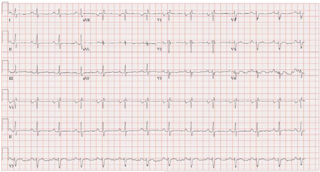
Figure 1: Electrocardiogram (ECG) demonstrating normal sinus rhythm with left atrial enlargement, rightward QRS axis, and incomplete right bundle branch block.
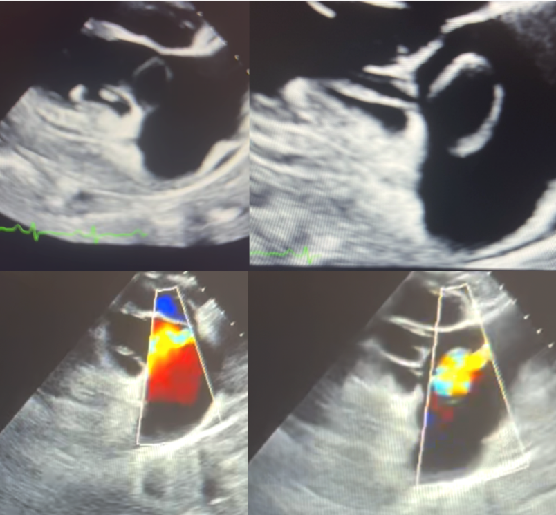
Figure 2: Transesophageal echocardiographic evaluation of the tricuspid valve. The left panes depict diastole, and the right panes depict systole; corresponding color Doppler imaging is shown in the lower panels. A cystic-appearing structure adjacent to the tricuspid valve annulus is noted, with color flow observed during both systole and diastole—findings suggestive of an aorto-atrial shunt from a ruptured sinus of Valsalva aneurysm.
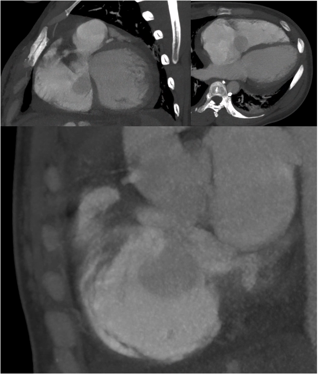
Figure 3: Nongated chest computed tomography angiography (CTA). Axial and coronal views demonstrate a prominent outpouching from the noncoronary sinus of Valsalva projecting into the right atrium, consistent with aneurysmal rupture. A region of swirling contrast with similar attenuation to the ascending aorta is visualized within the right atrium, indicative of a significant left-to-right shunt. Although limited by motion artifact and non-gated acquisition, the study provided key diagnostic clues supporting the diagnosis of ruptured sinus of Valsalva aneurysm (SOVAR).
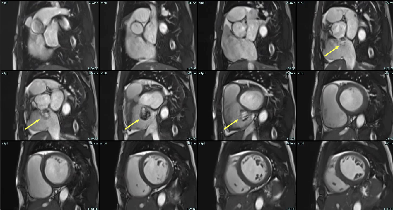
Figure 4: Cardiac magnetic resonance (CMR) short-axis steady-state free precession (SSFP) cine stack. Images demonstrate severe biventricular enlargement and a prominent cystic structure adjacent to the aortic root, consistent with a ruptured noncoronary sinus of Valsalva aneurysm projecting into the right atrium - yellow arrows highlight the site of rupture. The aneurysmal structure is contiguous with the aortic root and demonstrates dynamic filling, supporting the presence of an aorto-atrial communication.
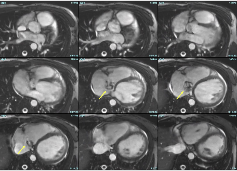
Figure 5: Axial steady-state free precession (SSFP) cine images from cardiac magnetic resonance (CMR), illustrating dynamic visualization of the ruptured sinus of Valsalva aneurysm. Yellow arrows denote the aneurysmal outpouching arising from the noncoronary sinus and extending into the right atrium. The signal variation and contour changes across frames reflect systolic-diastolic flow and deformation of the aneurysm, confirming an aorto-atrial communication. These images complement the short-axis stack in Figure 4 and emphasize the utility of multiplanar CMR imaging in localizing and characterizing shunt lesions.
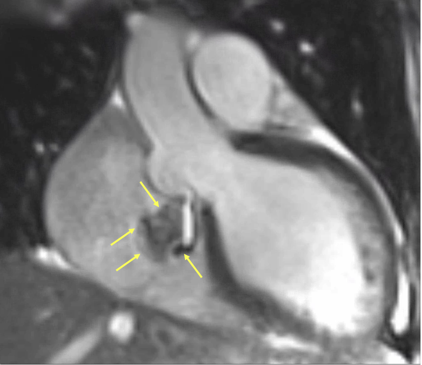
Figure 6: Magnified cardiac magnetic resonance (CMR) steady-state free precession (SSFP) cine still highlighting the ruptured sinus of Valsalva aneurysm. Yellow arrows delineate the borders of the aneurysmal sac arising from the noncoronary sinus and its direct extension into the right atrium. The swirling signal void within the aneurysm represents turbulent high-velocity shunting flow. This frame captures the anatomical and functional features of the rupture with high clarity, confirming an aorto-atrial communication and supporting the diagnosis suggested in Figures 4 and 5.
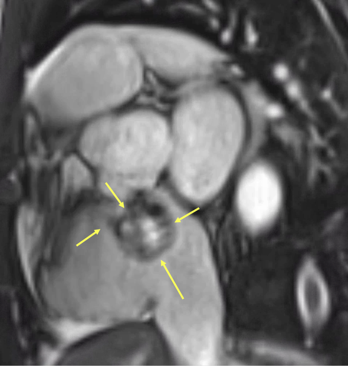
Figure 7: Cardiac magnetic resonance (CMR) short-axis steady-state free precession (SSFP) image at the level of the aortic root. Yellow arrows indicate the aneurysmal dilation arising from the noncoronary sinus of Valsalva, with a focal rupture site projecting into the right atrium. The central region demonstrates signal void consistent with turbulent high-velocity flow through the defect. This cross-sectional view confirms the origin and trajectory of the shunt, complementing the sagittal and axial projections shown in Figures 4–6.

Figure 8: Three-dimensional multiplanar reconstruction (3D MPR) from cardiac magnetic resonance imaging. Sequential static slices demonstrate an aneurysmal outpouching arising from the noncoronary sinus of Valsalva and projecting into the right atrium. The reconstruction highlights the anatomical morphology, orientation, and continuity of the rupture tract, offering precise localization of the aorto-atrial communication. This static dataset complements dynamic imaging by providing high spatial resolution for pre-surgical planning and anatomical delineation.
The pulmonary-to-systemic flow ratio (Qp:Qs), calculated using phase-contrast measurements at the ascending aorta and main pulmonary artery, was markedly elevated at 2.7–2.73 (Table 1), consistent with a significant left-to-right shunt. CMR also demonstrated severe biventricular chamber enlargement. Left ventricular ejection fraction was moderately reduced, while right ventricular ejection fraction was mildly reduced at 41% (Table 2). The tricuspid valve leaflets appeared structurally normal, with no evidence of stenosis. Mild tricuspid regurgitation was present, with a regurgitant fraction of approximately 8% on flow analysis. In contrast, moderate mitral regurgitation was observed, with a regurgitant fraction estimated at 38%, attributed to functional annular dilation.

Table 1: Biventricular volumetric and functional assessment by cardiac magnetic resonance imaging. The patient demonstrated severe biventricular chamber dilation with mildly to moderately reduced ejection fractions. Left ventricular (LV) and right ventricular (RV) cardiac outputs were markedly elevated, consistent with a high-output state likely driven by significant left-to-right shunting through the ruptured sinus of Valsalva aneurysm.

Table 2: Flow quantification by cardiac magnetic resonance (CMR) phase-contrast imaging. Main pulmonary artery (mPA) flow significantly exceeded systemic flow through the ascending aorta, consistent with a substantial left-to-right shunt. Systemic output was confirmed by summing flows through the superior vena cava (SVC) and descending aorta (dAo), yielding a Qp:Qs ratio of approximately 2.7.
We present a rare case of a peripartum ruptured sinus of Valsalva aneurysm in a patient who presented for the evaluation of progressive dyspnea, lower extremity edema, and concomitant occasional chest pain. Initial laboratory data, chest imaging, and physical examination all supported the diagnosis of acutely exacerbated heart failure with biventricular dilatation, pulmonary hypertension, and raising concerns for peripartum cardiomyopathy. CMR was pivotal in demonstrating the correct diagnosis of ruptured sinus of Valsalva with significant left to right shunt.
The patient was initiated on intravenous diuretics and guideline-directed medical therapy, including sacubitril-valsartan and dapagliflozin (Farxiga). In addition, iron supplementation was started for the treatment of anemia. She tolerated medical therapy well, with rapid improvement and eventual resolution of her symptoms. The patient remained hemodynamically and clinically stable, with complete resolution of her presenting symptoms and complaints. Cardiothoracic surgery was consulted for surgical evaluation.
Sinus of Valsalva aneurysm rupture into the right cardiac chambers is a rare congenital or acquired condition associated with significant morbidity and mortality (Weinreich et al., 2015). Its prevalence is estimated at 0.09% in the general population (Hope, 1842), most commonly involving the right coronary sinus, followed by the noncoronary sinus. The condition is more prevalent in men (4:1) and is significantly more common in individuals of Asian descent—up to five times higher than average—likely due to congenital predisposition (Feldman & Roman, 2006). Congenital causes include connective tissue disorders such as Marfan syndrome and Ehlers-Danlos syndrome. Acquired etiologies include trauma, atherosclerosis, infective endocarditis, iatrogenic injury, and syphilis. In our case, the aneurysmal rupture occurred in the peripartum period in a young woman without significant predisposing conditions—an extremely rare presentation with few documented cases in the literature (Huang et al., 2023; Latzman et al., 2006; Pamulapati et al., 1991).
The clinical presentation of SOVAR depends on several factors, including underlying comorbidities, the sinus involved, and the size of the defect. Acute rupture of a large aneurysm is associated with the worst prognosis, often presenting with sudden dyspnea, severe chest pain, and potential hemodynamic collapse. In contrast, a smaller, more gradual rupture may result in chronic, progressive symptoms that are more tolerable (Feldman & Roman, 2006). Despite its variable presentation, mortality remains high due to its elusive nature, underscoring the importance of a prompt, multimodal diagnostic approach when suspected.
A prominent sinus of Valsalva aneurysm rupture adjacent to the tricuspid valve annulus may precipitate significant tricuspid regurgitation in addition to creating an aorto-atrial shunt. This defect can lead to malcoaptation of the tricuspid leaflets due to wind-sock prolapse and forced systolic valve opening from the high-velocity shunting jet. While transthoracic echocardiography remains a cornerstone of initial cardiac imaging—particularly in patients presenting with heart failure symptoms—it may be limited in characterizing complex structural lesions. In such cases, a multimodality imaging approach is essential for accurately delineating the extent of the defect, identifying involved structures, cardiac chamber dimensions, assessing the severity of valvulopathy, and quantifying the degree of shunting. Different cases, such as in ours, may present with a cystic appearing lesion adjacent to the tricuspid valve demonstrating color flow. These parameters carry important diagnostic and prognostic value and are critical for guiding management.
Cardiac computed tomography angiography can also be a valuable tool in the diagnosis and management of sinus of Valsalva aneurysm rupture. This patient population is typically young and often at low to intermediate risk for obstructive coronary artery disease. In such cases, cCTA can reliably exclude coronary disease, potentially avoiding the need for invasive coronary angiography—particularly when the involved sinus corresponds to the origin of a coronary artery. Moreover, engagement of the coronary ostia during angiography may be technically challenging due to anatomical distortion and structural injury. While surgical repair remains the standard of care, emerging transcatheter, nonsurgical alternatives have been proposed (Latzman et al., 2006; Weinreich et al., 2015). In this context, cCTA can serve as an accurate tool for preprocedural planning. In our patient, SOVAR evaluation with chest CTA was partially limited by motion artifact at the level of the aortic root and suboptimal bolus timing for aortic evaluation. Nevertheless, it provided key diagnostic clues. Notably, there was evidence of negative contrast and swirling flow within the right atrium, largely isointense to the ascending aorta, consistent with a left-to-right shunt. Additionally, an aneurysmal outpouching of the non-coronary sinus projecting into the right atrium was clearly visualized.
Cardiac magnetic resonance imaging has been extensively validated for the evaluation of sinus of Valsalva aneurysm rupture. It enables precise quantification of cardiac chamber volumes, detailed assessment of valvular anatomy and function, and accurate measurement of flow parameters, including the pulmonary-to-systemic flow ratio (Qp:Qs). In addition to standard flow measurements at the aortic and pulmonary valves, CMR flow assessment can be cross-validated using alternative sites such as the descending aorta and superior vena cava. These secondary measurements provide internal consistency checks that enhance confidence in systemic and pulmonary flow quantification. In our case, systemic flow measurements obtained at the ascending aorta were concordant with those derived from the descending aorta and superior vena cava, thereby confirming the reliability of the findings and supporting the presence of a significant left-to-right shunt. The estimated Qp:Qs was approximately 2.7. Unfortunately, we were unable to obtain flow measurements at the left ventricular outflow tract or directly across the aortic valve. Such data would have enabled a more direct quantification of shunt volume and a more precise assessment of aortic regurgitation. This represents a learning opportunity for future cases involving similarly atypical presentations.
Furthermore, CINE imaging, particularly in non-standard planes tailored to individual anatomy, proved effective in visualizing diastolic flow into the receiving chamber, a key feature that helps distinguish shunt physiology from atrioventricular valve regurgitation. Additionally, the presence of sustained backward flow in the descending aorta has been previously correlated with significant aortic insufficiency. In our case, we postulate that this represents diastolic flow into the right atrium. This multimodal approach is especially valuable in evaluating complex structural lesions or in cases where initial echocardiographic findings are inconclusive. While right and left stroke volume measurements appeared relatively concordant, interpretation remains challenging. Nonetheless, the findings help exclude a pure intracardiac shunt such as a patent foramen ovale, atrial septal defect, or ventricular septal defect. However, these values must still be interpreted cautiously, given the volume overload imposed on one chamber and the potential presence of hemodynamically significant regurgitant valvular lesions.
An interesting finding in our patient’s case was the cystic appearance of the sinus of Valsalva aneurysm, which exhibited areas of signal loss on cardiac magnetic resonance imaging. Signal loss in CMR can arise due to several factors, including motion artifacts, flow effects, susceptibility artifacts, and intrinsic tissue properties. Given these potential causes, it is essential to tailor the interpretation to the specifics of each case. We hypothesize that signal loss is due to motion artifacts and turbulent blood flow, causing a void in the high-velocity shunting region. It is also important to note that certain pulse sequences and modifications in acquisition parameters can either exacerbate or mitigate signal loss. For instance, gradient echo sequences, while effective at minimizing some types of artifacts, are more susceptible to a loss of signal-to-noise ratio, making the choice of imaging parameters critical for accurate assessment and diagnosis. At times, a combination of different acquisition sequences and imaging parameters, rather than an either-or approach, should be considered to achieve a comprehensive and accurate evaluation.
It is crucial to exclude infective endocarditis as part of the differential diagnosis in patients presenting with sinus of Valsalva aneurysm rupture. First, infective endocarditis may serve as the underlying etiology, particularly in acquired cases where bacterial infection leads to focal destruction and weakening of the aortic wall or sinus. Second, its presence significantly increases the risk of morbidity and mortality due to complications such as septic embolization, abscess formation, valvular destruction, and systemic sepsis. The clinical presentation of SOVAR can closely resemble that of endocarditis-associated rupture, with both potentially manifesting as acute or progressive heart failure. However, infective endocarditis may also present with constitutional symptoms—such as fever, chills, malaise, and weight loss—which can provide important diagnostic clues. In our patient, there were no indications that the presentation was consistent with infective endocarditis, as she did not exhibit fever, chills, or other systemic signs of infection. Recognizing these features is critical, as the presence of infection would fundamentally alter both the urgency and nature of medical and surgical management. Prompt identification through blood cultures and transesophageal echocardiography is essential to guide appropriate therapy and improve outcomes.
In this case, the cardiac magnetic resonance was instrumental in achieving the correct diagnosis of a ruptured sinus of Valsalva aneurysm. While other imaging modalities, including transthoracic echocardiography and cardiac computed tomography angiography, accurately described the aneurysm, they were unable to fully characterize the ruptured component or qualitatively and quantitatively assess the degree of shunting. The importance of CMR in such cases cannot be overstated; it allows for precise chamber volume quantification, accurate dimensional evaluation, and is considered the gold standard for biventricular ejection fraction estimation. CMR also provides unparalleled shunt quantification, which is critical in evaluating the severity of left-to-right shunting. Additionally, the ability to obtain 3D imaging and CINE sequences offers valuable insights that are difficult, if not impossible, to capture through TTE or transesophageal echocardiography. Although cardiac CTA can play a role in diagnosing such conditions, its protocol is limited to imaging only one cardiac cycle or a portion of it, making it less effective in capturing dynamic aspects of aneurysms and shunts. Ultimately, CMR proved to be essential in diagnosing the ruptured sinus of Valsalva aneurysm and guiding the management and therapeutic decisions of the patient.
This case highlights a rare but life-threatening presentation of ruptured sinus of Valsalva aneurysm in the peripartum period, initially masquerading as peripartum cardiomyopathy. The diagnostic complexity underscores the need for a high index of suspicion and a systematic, multimodality imaging approach. Cardiac magnetic resonance imaging was instrumental in accurately identifying the rupture, quantifying the left-to-right shunt, and excluding other structural or ischemic etiologies. This case reinforces the critical role of advanced imaging in evaluating postpartum patients with unexplained dyspnea and emphasizes the importance of considering structural heart disease—even in the absence of classic risk factors or overt signs. Early diagnosis and tailored therapy can lead to favorable clinical outcomes, even in uncommon and deceptive presentations such as this.
Clearly Auctoresonline and particularly Psychology and Mental Health Care Journal is dedicated to improving health care services for individuals and populations. The editorial boards' ability to efficiently recognize and share the global importance of health literacy with a variety of stakeholders. Auctoresonline publishing platform can be used to facilitate of optimal client-based services and should be added to health care professionals' repertoire of evidence-based health care resources.

Journal of Clinical Cardiology and Cardiovascular Intervention The submission and review process was adequate. However I think that the publication total value should have been enlightened in early fases. Thank you for all.

Journal of Women Health Care and Issues By the present mail, I want to say thank to you and tour colleagues for facilitating my published article. Specially thank you for the peer review process, support from the editorial office. I appreciate positively the quality of your journal.

Journal of Clinical Research and Reports I would be very delighted to submit my testimonial regarding the reviewer board and the editorial office. The reviewer board were accurate and helpful regarding any modifications for my manuscript. And the editorial office were very helpful and supportive in contacting and monitoring with any update and offering help. It was my pleasure to contribute with your promising Journal and I am looking forward for more collaboration.

We would like to thank the Journal of Thoracic Disease and Cardiothoracic Surgery because of the services they provided us for our articles. The peer-review process was done in a very excellent time manner, and the opinions of the reviewers helped us to improve our manuscript further. The editorial office had an outstanding correspondence with us and guided us in many ways. During a hard time of the pandemic that is affecting every one of us tremendously, the editorial office helped us make everything easier for publishing scientific work. Hope for a more scientific relationship with your Journal.

The peer-review process which consisted high quality queries on the paper. I did answer six reviewers’ questions and comments before the paper was accepted. The support from the editorial office is excellent.

Journal of Neuroscience and Neurological Surgery. I had the experience of publishing a research article recently. The whole process was simple from submission to publication. The reviewers made specific and valuable recommendations and corrections that improved the quality of my publication. I strongly recommend this Journal.

Dr. Katarzyna Byczkowska My testimonial covering: "The peer review process is quick and effective. The support from the editorial office is very professional and friendly. Quality of the Clinical Cardiology and Cardiovascular Interventions is scientific and publishes ground-breaking research on cardiology that is useful for other professionals in the field.

Thank you most sincerely, with regard to the support you have given in relation to the reviewing process and the processing of my article entitled "Large Cell Neuroendocrine Carcinoma of The Prostate Gland: A Review and Update" for publication in your esteemed Journal, Journal of Cancer Research and Cellular Therapeutics". The editorial team has been very supportive.

Testimony of Journal of Clinical Otorhinolaryngology: work with your Reviews has been a educational and constructive experience. The editorial office were very helpful and supportive. It was a pleasure to contribute to your Journal.

Dr. Bernard Terkimbi Utoo, I am happy to publish my scientific work in Journal of Women Health Care and Issues (JWHCI). The manuscript submission was seamless and peer review process was top notch. I was amazed that 4 reviewers worked on the manuscript which made it a highly technical, standard and excellent quality paper. I appreciate the format and consideration for the APC as well as the speed of publication. It is my pleasure to continue with this scientific relationship with the esteem JWHCI.

This is an acknowledgment for peer reviewers, editorial board of Journal of Clinical Research and Reports. They show a lot of consideration for us as publishers for our research article “Evaluation of the different factors associated with side effects of COVID-19 vaccination on medical students, Mutah university, Al-Karak, Jordan”, in a very professional and easy way. This journal is one of outstanding medical journal.

Dear Hao Jiang, to Journal of Nutrition and Food Processing We greatly appreciate the efficient, professional and rapid processing of our paper by your team. If there is anything else we should do, please do not hesitate to let us know. On behalf of my co-authors, we would like to express our great appreciation to editor and reviewers.

As an author who has recently published in the journal "Brain and Neurological Disorders". I am delighted to provide a testimonial on the peer review process, editorial office support, and the overall quality of the journal. The peer review process at Brain and Neurological Disorders is rigorous and meticulous, ensuring that only high-quality, evidence-based research is published. The reviewers are experts in their fields, and their comments and suggestions were constructive and helped improve the quality of my manuscript. The review process was timely and efficient, with clear communication from the editorial office at each stage. The support from the editorial office was exceptional throughout the entire process. The editorial staff was responsive, professional, and always willing to help. They provided valuable guidance on formatting, structure, and ethical considerations, making the submission process seamless. Moreover, they kept me informed about the status of my manuscript and provided timely updates, which made the process less stressful. The journal Brain and Neurological Disorders is of the highest quality, with a strong focus on publishing cutting-edge research in the field of neurology. The articles published in this journal are well-researched, rigorously peer-reviewed, and written by experts in the field. The journal maintains high standards, ensuring that readers are provided with the most up-to-date and reliable information on brain and neurological disorders. In conclusion, I had a wonderful experience publishing in Brain and Neurological Disorders. The peer review process was thorough, the editorial office provided exceptional support, and the journal's quality is second to none. I would highly recommend this journal to any researcher working in the field of neurology and brain disorders.
Dear Agrippa Hilda, Journal of Neuroscience and Neurological Surgery, Editorial Coordinator, I trust this message finds you well. I want to extend my appreciation for considering my article for publication in your esteemed journal. I am pleased to provide a testimonial regarding the peer review process and the support received from your editorial office. The peer review process for my paper was carried out in a highly professional and thorough manner. The feedback and comments provided by the authors were constructive and very useful in improving the quality of the manuscript. This rigorous assessment process undoubtedly contributes to the high standards maintained by your journal.

International Journal of Clinical Case Reports and Reviews. I strongly recommend to consider submitting your work to this high-quality journal. The support and availability of the Editorial staff is outstanding and the review process was both efficient and rigorous.

Thank you very much for publishing my Research Article titled “Comparing Treatment Outcome Of Allergic Rhinitis Patients After Using Fluticasone Nasal Spray And Nasal Douching" in the Journal of Clinical Otorhinolaryngology. As Medical Professionals we are immensely benefited from study of various informative Articles and Papers published in this high quality Journal. I look forward to enriching my knowledge by regular study of the Journal and contribute my future work in the field of ENT through the Journal for use by the medical fraternity. The support from the Editorial office was excellent and very prompt. I also welcome the comments received from the readers of my Research Article.

Dear Erica Kelsey, Editorial Coordinator of Cancer Research and Cellular Therapeutics Our team is very satisfied with the processing of our paper by your journal. That was fast, efficient, rigorous, but without unnecessary complications. We appreciated the very short time between the submission of the paper and its publication on line on your site.

I am very glad to say that the peer review process is very successful and fast and support from the Editorial Office. Therefore, I would like to continue our scientific relationship for a long time. And I especially thank you for your kindly attention towards my article. Have a good day!

"We recently published an article entitled “Influence of beta-Cyclodextrins upon the Degradation of Carbofuran Derivatives under Alkaline Conditions" in the Journal of “Pesticides and Biofertilizers” to show that the cyclodextrins protect the carbamates increasing their half-life time in the presence of basic conditions This will be very helpful to understand carbofuran behaviour in the analytical, agro-environmental and food areas. We greatly appreciated the interaction with the editor and the editorial team; we were particularly well accompanied during the course of the revision process, since all various steps towards publication were short and without delay".

I would like to express my gratitude towards you process of article review and submission. I found this to be very fair and expedient. Your follow up has been excellent. I have many publications in national and international journal and your process has been one of the best so far. Keep up the great work.

We are grateful for this opportunity to provide a glowing recommendation to the Journal of Psychiatry and Psychotherapy. We found that the editorial team were very supportive, helpful, kept us abreast of timelines and over all very professional in nature. The peer review process was rigorous, efficient and constructive that really enhanced our article submission. The experience with this journal remains one of our best ever and we look forward to providing future submissions in the near future.

I am very pleased to serve as EBM of the journal, I hope many years of my experience in stem cells can help the journal from one way or another. As we know, stem cells hold great potential for regenerative medicine, which are mostly used to promote the repair response of diseased, dysfunctional or injured tissue using stem cells or their derivatives. I think Stem Cell Research and Therapeutics International is a great platform to publish and share the understanding towards the biology and translational or clinical application of stem cells.

I would like to give my testimony in the support I have got by the peer review process and to support the editorial office where they were of asset to support young author like me to be encouraged to publish their work in your respected journal and globalize and share knowledge across the globe. I really give my great gratitude to your journal and the peer review including the editorial office.

I am delighted to publish our manuscript entitled "A Perspective on Cocaine Induced Stroke - Its Mechanisms and Management" in the Journal of Neuroscience and Neurological Surgery. The peer review process, support from the editorial office, and quality of the journal are excellent. The manuscripts published are of high quality and of excellent scientific value. I recommend this journal very much to colleagues.

Dr.Tania Muñoz, My experience as researcher and author of a review article in The Journal Clinical Cardiology and Interventions has been very enriching and stimulating. The editorial team is excellent, performs its work with absolute responsibility and delivery. They are proactive, dynamic and receptive to all proposals. Supporting at all times the vast universe of authors who choose them as an option for publication. The team of review specialists, members of the editorial board, are brilliant professionals, with remarkable performance in medical research and scientific methodology. Together they form a frontline team that consolidates the JCCI as a magnificent option for the publication and review of high-level medical articles and broad collective interest. I am honored to be able to share my review article and open to receive all your comments.

“The peer review process of JPMHC is quick and effective. Authors are benefited by good and professional reviewers with huge experience in the field of psychology and mental health. The support from the editorial office is very professional. People to contact to are friendly and happy to help and assist any query authors might have. Quality of the Journal is scientific and publishes ground-breaking research on mental health that is useful for other professionals in the field”.

Dear editorial department: On behalf of our team, I hereby certify the reliability and superiority of the International Journal of Clinical Case Reports and Reviews in the peer review process, editorial support, and journal quality. Firstly, the peer review process of the International Journal of Clinical Case Reports and Reviews is rigorous, fair, transparent, fast, and of high quality. The editorial department invites experts from relevant fields as anonymous reviewers to review all submitted manuscripts. These experts have rich academic backgrounds and experience, and can accurately evaluate the academic quality, originality, and suitability of manuscripts. The editorial department is committed to ensuring the rigor of the peer review process, while also making every effort to ensure a fast review cycle to meet the needs of authors and the academic community. Secondly, the editorial team of the International Journal of Clinical Case Reports and Reviews is composed of a group of senior scholars and professionals with rich experience and professional knowledge in related fields. The editorial department is committed to assisting authors in improving their manuscripts, ensuring their academic accuracy, clarity, and completeness. Editors actively collaborate with authors, providing useful suggestions and feedback to promote the improvement and development of the manuscript. We believe that the support of the editorial department is one of the key factors in ensuring the quality of the journal. Finally, the International Journal of Clinical Case Reports and Reviews is renowned for its high- quality articles and strict academic standards. The editorial department is committed to publishing innovative and academically valuable research results to promote the development and progress of related fields. The International Journal of Clinical Case Reports and Reviews is reasonably priced and ensures excellent service and quality ratio, allowing authors to obtain high-level academic publishing opportunities in an affordable manner. I hereby solemnly declare that the International Journal of Clinical Case Reports and Reviews has a high level of credibility and superiority in terms of peer review process, editorial support, reasonable fees, and journal quality. Sincerely, Rui Tao.

Clinical Cardiology and Cardiovascular Interventions I testity the covering of the peer review process, support from the editorial office, and quality of the journal.

Clinical Cardiology and Cardiovascular Interventions, we deeply appreciate the interest shown in our work and its publication. It has been a true pleasure to collaborate with you. The peer review process, as well as the support provided by the editorial office, have been exceptional, and the quality of the journal is very high, which was a determining factor in our decision to publish with you.

The peer reviewers process is quick and effective, the supports from editorial office is excellent, the quality of journal is high. I would like to collabroate with Internatioanl journal of Clinical Case Reports and Reviews journal clinically in the future time.

Clinical Cardiology and Cardiovascular Interventions, I would like to express my sincerest gratitude for the trust placed in our team for the publication in your journal. It has been a true pleasure to collaborate with you on this project. I am pleased to inform you that both the peer review process and the attention from the editorial coordination have been excellent. Your team has worked with dedication and professionalism to ensure that your publication meets the highest standards of quality. We are confident that this collaboration will result in mutual success, and we are eager to see the fruits of this shared effort.

Dear Dr. Jessica Magne, Editorial Coordinator 0f Clinical Cardiology and Cardiovascular Interventions, I hope this message finds you well. I want to express my utmost gratitude for your excellent work and for the dedication and speed in the publication process of my article titled "Navigating Innovation: Qualitative Insights on Using Technology for Health Education in Acute Coronary Syndrome Patients." I am very satisfied with the peer review process, the support from the editorial office, and the quality of the journal. I hope we can maintain our scientific relationship in the long term.

Dear Monica Gissare, - Editorial Coordinator of Nutrition and Food Processing. ¨My testimony with you is truly professional, with a positive response regarding the follow-up of the article and its review, you took into account my qualities and the importance of the topic¨.

Dear Dr. Jessica Magne, Editorial Coordinator 0f Clinical Cardiology and Cardiovascular Interventions, The review process for the article “The Handling of Anti-aggregants and Anticoagulants in the Oncologic Heart Patient Submitted to Surgery” was extremely rigorous and detailed. From the initial submission to the final acceptance, the editorial team at the “Journal of Clinical Cardiology and Cardiovascular Interventions” demonstrated a high level of professionalism and dedication. The reviewers provided constructive and detailed feedback, which was essential for improving the quality of our work. Communication was always clear and efficient, ensuring that all our questions were promptly addressed. The quality of the “Journal of Clinical Cardiology and Cardiovascular Interventions” is undeniable. It is a peer-reviewed, open-access publication dedicated exclusively to disseminating high-quality research in the field of clinical cardiology and cardiovascular interventions. The journal's impact factor is currently under evaluation, and it is indexed in reputable databases, which further reinforces its credibility and relevance in the scientific field. I highly recommend this journal to researchers looking for a reputable platform to publish their studies.

Dear Editorial Coordinator of the Journal of Nutrition and Food Processing! "I would like to thank the Journal of Nutrition and Food Processing for including and publishing my article. The peer review process was very quick, movement and precise. The Editorial Board has done an extremely conscientious job with much help, valuable comments and advices. I find the journal very valuable from a professional point of view, thank you very much for allowing me to be part of it and I would like to participate in the future!”

Dealing with The Journal of Neurology and Neurological Surgery was very smooth and comprehensive. The office staff took time to address my needs and the response from editors and the office was prompt and fair. I certainly hope to publish with this journal again.Their professionalism is apparent and more than satisfactory. Susan Weiner

My Testimonial Covering as fellowing: Lin-Show Chin. The peer reviewers process is quick and effective, the supports from editorial office is excellent, the quality of journal is high. I would like to collabroate with Internatioanl journal of Clinical Case Reports and Reviews.

My experience publishing in Psychology and Mental Health Care was exceptional. The peer review process was rigorous and constructive, with reviewers providing valuable insights that helped enhance the quality of our work. The editorial team was highly supportive and responsive, making the submission process smooth and efficient. The journal's commitment to high standards and academic rigor makes it a respected platform for quality research. I am grateful for the opportunity to publish in such a reputable journal.

My experience publishing in International Journal of Clinical Case Reports and Reviews was exceptional. I Come forth to Provide a Testimonial Covering the Peer Review Process and the editorial office for the Professional and Impartial Evaluation of the Manuscript.

I would like to offer my testimony in the support. I have received through the peer review process and support the editorial office where they are to support young authors like me, encourage them to publish their work in your esteemed journals, and globalize and share knowledge globally. I really appreciate your journal, peer review, and editorial office.

Dear Agrippa Hilda- Editorial Coordinator of Journal of Neuroscience and Neurological Surgery, "The peer review process was very quick and of high quality, which can also be seen in the articles in the journal. The collaboration with the editorial office was very good."

I would like to express my sincere gratitude for the support and efficiency provided by the editorial office throughout the publication process of my article, “Delayed Vulvar Metastases from Rectal Carcinoma: A Case Report.” I greatly appreciate the assistance and guidance I received from your team, which made the entire process smooth and efficient. The peer review process was thorough and constructive, contributing to the overall quality of the final article. I am very grateful for the high level of professionalism and commitment shown by the editorial staff, and I look forward to maintaining a long-term collaboration with the International Journal of Clinical Case Reports and Reviews.

To Dear Erin Aust, I would like to express my heartfelt appreciation for the opportunity to have my work published in this esteemed journal. The entire publication process was smooth and well-organized, and I am extremely satisfied with the final result. The Editorial Team demonstrated the utmost professionalism, providing prompt and insightful feedback throughout the review process. Their clear communication and constructive suggestions were invaluable in enhancing my manuscript, and their meticulous attention to detail and dedication to quality are truly commendable. Additionally, the support from the Editorial Office was exceptional. From the initial submission to the final publication, I was guided through every step of the process with great care and professionalism. The team's responsiveness and assistance made the entire experience both easy and stress-free. I am also deeply impressed by the quality and reputation of the journal. It is an honor to have my research featured in such a respected publication, and I am confident that it will make a meaningful contribution to the field.

"I am grateful for the opportunity of contributing to [International Journal of Clinical Case Reports and Reviews] and for the rigorous review process that enhances the quality of research published in your esteemed journal. I sincerely appreciate the time and effort of your team who have dedicatedly helped me in improvising changes and modifying my manuscript. The insightful comments and constructive feedback provided have been invaluable in refining and strengthening my work".

I thank the ‘Journal of Clinical Research and Reports’ for accepting this article for publication. This is a rigorously peer reviewed journal which is on all major global scientific data bases. I note the review process was prompt, thorough and professionally critical. It gave us an insight into a number of important scientific/statistical issues. The review prompted us to review the relevant literature again and look at the limitations of the study. The peer reviewers were open, clear in the instructions and the editorial team was very prompt in their communication. This journal certainly publishes quality research articles. I would recommend the journal for any future publications.

Dear Jessica Magne, with gratitude for the joint work. Fast process of receiving and processing the submitted scientific materials in “Clinical Cardiology and Cardiovascular Interventions”. High level of competence of the editors with clear and correct recommendations and ideas for enriching the article.

We found the peer review process quick and positive in its input. The support from the editorial officer has been very agile, always with the intention of improving the article and taking into account our subsequent corrections.

My article, titled 'No Way Out of the Smartphone Epidemic Without Considering the Insights of Brain Research,' has been republished in the International Journal of Clinical Case Reports and Reviews. The review process was seamless and professional, with the editors being both friendly and supportive. I am deeply grateful for their efforts.

To Dear Erin Aust – Editorial Coordinator of Journal of General Medicine and Clinical Practice! I declare that I am absolutely satisfied with your work carried out with great competence in following the manuscript during the various stages from its receipt, during the revision process to the final acceptance for publication. Thank Prof. Elvira Farina

Dear Jessica, and the super professional team of the ‘Clinical Cardiology and Cardiovascular Interventions’ I am sincerely grateful to the coordinated work of the journal team for the no problem with the submission of my manuscript: “Cardiometabolic Disorders in A Pregnant Woman with Severe Preeclampsia on the Background of Morbid Obesity (Case Report).” The review process by 5 experts was fast, and the comments were professional, which made it more specific and academic, and the process of publication and presentation of the article was excellent. I recommend that my colleagues publish articles in this journal, and I am interested in further scientific cooperation. Sincerely and best wishes, Dr. Oleg Golyanovskiy.

Dear Ashley Rosa, Editorial Coordinator of the journal - Psychology and Mental Health Care. " The process of obtaining publication of my article in the Psychology and Mental Health Journal was positive in all areas. The peer review process resulted in a number of valuable comments, the editorial process was collaborative and timely, and the quality of this journal has been quickly noticed, resulting in alternative journals contacting me to publish with them." Warm regards, Susan Anne Smith, PhD. Australian Breastfeeding Association.

Dear Jessica Magne, Editorial Coordinator, Clinical Cardiology and Cardiovascular Interventions, Auctores Publishing LLC. I appreciate the journal (JCCI) editorial office support, the entire team leads were always ready to help, not only on technical front but also on thorough process. Also, I should thank dear reviewers’ attention to detail and creative approach to teach me and bring new insights by their comments. Surely, more discussions and introduction of other hemodynamic devices would provide better prevention and management of shock states. Your efforts and dedication in presenting educational materials in this journal are commendable. Best wishes from, Farahnaz Fallahian.

Dear Maria Emerson, Editorial Coordinator, International Journal of Clinical Case Reports and Reviews, Auctores Publishing LLC. I am delighted to have published our manuscript, "Acute Colonic Pseudo-Obstruction (ACPO): A rare but serious complication following caesarean section." I want to thank the editorial team, especially Maria Emerson, for their prompt review of the manuscript, quick responses to queries, and overall support. Yours sincerely Dr. Victor Olagundoye.

Dear Ashley Rosa, Editorial Coordinator, International Journal of Clinical Case Reports and Reviews. Many thanks for publishing this manuscript after I lost confidence the editors were most helpful, more than other journals Best wishes from, Susan Anne Smith, PhD. Australian Breastfeeding Association.

Dear Agrippa Hilda, Editorial Coordinator, Journal of Neuroscience and Neurological Surgery. The entire process including article submission, review, revision, and publication was extremely easy. The journal editor was prompt and helpful, and the reviewers contributed to the quality of the paper. Thank you so much! Eric Nussbaum, MD

Dr Hala Al Shaikh This is to acknowledge that the peer review process for the article ’ A Novel Gnrh1 Gene Mutation in Four Omani Male Siblings, Presentation and Management ’ sent to the International Journal of Clinical Case Reports and Reviews was quick and smooth. The editorial office was prompt with easy communication.

Dear Erin Aust, Editorial Coordinator, Journal of General Medicine and Clinical Practice. We are pleased to share our experience with the “Journal of General Medicine and Clinical Practice”, following the successful publication of our article. The peer review process was thorough and constructive, helping to improve the clarity and quality of the manuscript. We are especially thankful to Ms. Erin Aust, the Editorial Coordinator, for her prompt communication and continuous support throughout the process. Her professionalism ensured a smooth and efficient publication experience. The journal upholds high editorial standards, and we highly recommend it to fellow researchers seeking a credible platform for their work. Best wishes By, Dr. Rakhi Mishra.

Dear Jessica Magne, Editorial Coordinator, Clinical Cardiology and Cardiovascular Interventions, Auctores Publishing LLC. The peer review process of the journal of Clinical Cardiology and Cardiovascular Interventions was excellent and fast, as was the support of the editorial office and the quality of the journal. Kind regards Walter F. Riesen Prof. Dr. Dr. h.c. Walter F. Riesen.

Dear Ashley Rosa, Editorial Coordinator, International Journal of Clinical Case Reports and Reviews, Auctores Publishing LLC. Thank you for publishing our article, Exploring Clozapine's Efficacy in Managing Aggression: A Multiple Single-Case Study in Forensic Psychiatry in the international journal of clinical case reports and reviews. We found the peer review process very professional and efficient. The comments were constructive, and the whole process was efficient. On behalf of the co-authors, I would like to thank you for publishing this article. With regards, Dr. Jelle R. Lettinga.

Dear Clarissa Eric, Editorial Coordinator, Journal of Clinical Case Reports and Studies, I would like to express my deep admiration for the exceptional professionalism demonstrated by your journal. I am thoroughly impressed by the speed of the editorial process, the substantive and insightful reviews, and the meticulous preparation of the manuscript for publication. Additionally, I greatly appreciate the courteous and immediate responses from your editorial office to all my inquiries. Best Regards, Dariusz Ziora

Dear Chrystine Mejia, Editorial Coordinator, Journal of Neurodegeneration and Neurorehabilitation, Auctores Publishing LLC, We would like to thank the editorial team for the smooth and high-quality communication leading up to the publication of our article in the Journal of Neurodegeneration and Neurorehabilitation. The reviewers have extensive knowledge in the field, and their relevant questions helped to add value to our publication. Kind regards, Dr. Ravi Shrivastava.

Dear Clarissa Eric, Editorial Coordinator, Journal of Clinical Case Reports and Studies, Auctores Publishing LLC, USA Office: +1-(302)-520-2644. I would like to express my sincere appreciation for the efficient and professional handling of my case report by the ‘Journal of Clinical Case Reports and Studies’. The peer review process was not only fast but also highly constructive—the reviewers’ comments were clear, relevant, and greatly helped me improve the quality and clarity of my manuscript. I also received excellent support from the editorial office throughout the process. Communication was smooth and timely, and I felt well guided at every stage, from submission to publication. The overall quality and rigor of the journal are truly commendable. I am pleased to have published my work with Journal of Clinical Case Reports and Studies, and I look forward to future opportunities for collaboration. Sincerely, Aline Tollet, UCLouvain.

Dear Ms. Mayra Duenas, Editorial Coordinator, International Journal of Clinical Case Reports and Reviews. “The International Journal of Clinical Case Reports and Reviews represented the “ideal house” to share with the research community a first experience with the use of the Simeox device for speech rehabilitation. High scientific reputation and attractive website communication were first determinants for the selection of this Journal, and the following submission process exceeded expectations: fast but highly professional peer review, great support by the editorial office, elegant graphic layout. Exactly what a dynamic research team - also composed by allied professionals - needs!" From, Chiara Beccaluva, PT - Italy.
