AUCTORES
Globalize your Research

Case report | DOI: https://doi.org/10.31579/2578-8868/369
Head of Neurosurgical Department, Juaneda Miramar, Palma de Mallorca, Balearic Islands, Spain
*Corresponding Author: Gonçalo Januário, Juaneda Miramar, Neurosurgical Department, Palma de Mallorca, Balearic Islands, Spain.
Citation: Gonçalo Januário (2025), Duane Syndrome Combined with Intracranial Cisternal Lipoma and Syringomyelia, Case Report and a Review of Literature, J. Neuroscience and Neurological Surgery, 17(4); DOI:10.31579/2578-8868/369
Copyright: © 2025, Gonçalo Januário. This is an open-access article distributed under the terms of The Creative Commons Attribution License, which permits unrestricted use, distribution, and reproduction in any medium, provided the original author and source are credited
Received: 07 March 2025 | Accepted: 26 March 2025 | Published: 16 April 2025
Keywords: intracranial lipoma; quadrigeminal cistern; duane syndrome; syringomyelia; magnetic resonance imaging
Intracranial lipomas (IL) are very uncommon congenital malformations. Frequently are associated with various brain anomalies as agenesis or dysgenesis of the adjacent structures like corpus callosum or cortical dysplasia.Some IL are asymptomatic, incidentally discovered in brain imaging carried out to any other causes. The most common location is interhemispheric accounting for 45% of cases. In 20-25% they are located in ambient or quadrigeminal cisterns.Duane syndrome (DS) is a congenital type of strabismus. About 80-90% of cases are unilateral and the left eye is more commonly affected. Are there different types of DS and this pathology doesn’t always happen with other conditions, sometimes are associated with hearing disorders, Goldenhar syndrome, Malformations in spine or vertebrae. The patients with this disease may also present other eye-related conditions as cataract, microphthalmos, nystagmus.Differential Diagnosis for DS includes, Okihiro's syndrome, Goldenhar syndrome, Wildervanck syndrome, Moebius Syndrome, Holt-Oram Syndrome, Morning Glory Syndrome, Abducens Nerve Palsy, Brown Syndrome, Marcus Gunn Jaw Winking Syndrome, and Congenital Esotropia.We present a clinical case, very uncommon, in a 12 year old boy with Duane Syndrome that affects the left eye. The patient present also an IL located in the right quadrigeminal cistern. Incidentally, a dorsal syringomyelic lesion was discovered.All of them are uncommon pathologies, an intracranial lipoma with this location, Duane syndrome and thoracic syringomyelia. We did not find any case in the literature that describes the three pathologies in the same patient.
Intracranial lipoma (IL) is a congenital slow-growing, benign tumor made up of adipose tissue. Although they often remain undetected until later in life. Rokitansky was the first author to describe intracranial lipomas in 19th century, 1856. [1] These benign tumors represent less than 0.1% of all intracranial tumors. [2] The most common location of the intracranial lipomas is Interhemispheric accounting for 45% of cases. The other lesion’s locations were clustered in the quadrigeminal/superior cerebellar in 25%, suprasellar/interpeduncular in 14%, cerebellopontine angle in 9%, and sylvian cisterns in 5%. The IL were associated with brain malformations of varying degrees in 55% of cases. An interesting note for potential surgical cases, in 36% of the cases the intracranial vessels and nerves course through the lesions. [3]
They are congenital lesions composed of adipose tissue resulting from abnormal persistence as well as altered differentiation of the primitive embryonic meninges during the development of the subarachnoid cisterns. The altered differentiation of the meningeal tissue during embryogenesis is its most likely origin. This embryologic concept of the development of IL presents a theory to explain the high frequency of callosal and other brain hypoplasias associated with this benign lesion. [4]
Some IL are asymptomatic, incidentally discovered in brain imaging carried out to any other organic cause. Symptoms, if present, are associated with increased intracranial pressure, obstructive hydrocephalus, or seizures. They can manifest with headache, mental dysfunction, and cranial nerve deficits. [5,6,7] Clinical manifestations depends on the location of the lesion and mainly are asymptomatic, represent incidental lesions, discovered on Computed Tomography (CT) or Magnetic Resonance Imaging (MRI), performed for other reason such as head trauma or headaches. The typical findings in both imaging techniques are the presence of a lesion with appearance and characteristics compatible with fat. On CT scan, IL appear as a non-enhancing lesion with uniform fat density, in some cases may exist peripheral calcification. The CT and MRI features are often pathognomonic for intracranial lipomas.7The treatment are manly conservative, and surgical procedure is indicated in situations with cosmetic defects that require resection of the extracranial portion of the lipoma, obstructive hydrocephalus with indication for a cerebrospinal fluid (CSF) diversion procedure or ventriculostomy, uncontrollable seizures.[8] When indicated the resection could be extremely difficult and potentially risky and dangerous. Is especially related with the location of the mass and because the huge adhesion with surrounding brain parenchyma, neurological and vascular structures.[9] Duane retraction syndrome (DRS), also called Stilling–Turk–Duane syndrome, is a congenital eye movement abnormality that present a variable horizontal duction deficits, with narrowing of the palpebral fissure and globe retraction on attempted abduction, occasionally accompanied by upshoot or downshoot.[10] The DRS is a cause of all forms of strabismus in 5% of the cases, it was widely described in the literature as early as the 19th century.[11,12] In 1905 Alexander Duane published a series of 54 cases with DRS, did a description of the clinical features and resume the possible etiopathogenesis and management of this pathology. With the advent and development of neuroimaging, muscle electrophysiology and genetic analysis, there has been greater understanding of this form of strabismus, now considered a congenital cranial dysinnervation disorder (CCDD), giving better insights into the management of this challenging syndrome.[13]
We describe a clinical case of a 12-year-old male child, eutocic delivery with APGAR score of 9 in the first minute and 10 at 5 minutes. Was born with a weight of 3315 kg, a height of 48 cm, and a cranial circumference of 34 cm. Blood group O RH -, with negative direct Coombs. At delivery, a dermolipoma was observed in the left eye and 2 left preauricular appendages, patent external ear pavilions. Hemifacial microstomia (Goldenhar syndrome was subsequently ruled out). No other visible morphological alterations. Considering the identified alterations, he was referred and periodically evaluated by Neonatology, Pediatrics, Ophthalmology, Otorhinolaryngology, Neurology and later by Neurosurgery. In this context, an MRI was performed, where a left intraorbital lesion with fatty content was observed adjacent to the lateral wall of the eyeball and in front of the insertion of the lateral rectus muscle. Small dermoid cyst in the right peripontine cistern, hyperintensity in T1. At one month of age, a routine check-up revealed a head circumference of 45 cm with plagiocephaly, short neck, functional limitation of the left VI PC, eyelid asymmetry, blinking with myoclonus in both eyes when looking up. Occasional episodes lasting a few seconds. The patient has been monitored and periodically evaluated in the neonatal pediatrics department; he has shown active, normal muscle tone, positive suction-search, complete bilateral moro, positive palmoplantar pressure, positive galant test. Convergent strabismus of the left eye. An electroencephalogram was also performed, which showed no abnormalities and was reported as normal for his age. To rule out organic pathology, a cardiac ultrasound was performed: normal, with no cardiac symptoms ever present. A renal ultrasound was also performed, which showed no abnormalities. A genetic study was performed, showing karyotype 46XY, normal (Figure 1).

Figure 1: Karyotype normal, with a 46 XY.
During the follow-up at 8 months of age, the laboratory tests identified a heterozygous C677T mutation (MTHFR gene) and the thyroid study was normal. A coagulation disorder was identified with a homozygous coagulation factor XII mutation.
We highlight the evaluation in the Pediatrics consultation at one year of life, in which the patient presented free crawling, begins walking, social smile, does not point with the finger. Does not close hands in the midline. Poor imitation of movements. Responds to his name. Discrete left superior orbicularis muscle asymmetry. Dermolipoma in the left eye with
paresis of the VI PC. Convergent strabismus in the left eye. Normal ENT evaluation.
An brain MRI was performed during the same period that revealed a well-defined homogenous fat density seen in the midline along the falx cerebri at the level of the vertex. It measured 2.5 x 1.5 x 4 cm in maximum AP x TR x CC [anteroposterior x transverse x craniocaudal] dimensions, likely representing quadrigeminal cistern lipoma. Left intraorbital lesion with fatty content adjacent to the lateral wall of the eyeball. Both lesions showed no changes in dimension or density compared to previous MRIs.
The child’s condition remained stable there was no headache, dizziness, abnormal movements, or seizures. In 2018 repeat brain mri and the findings are substantially similar. Brain morphology within normality for age, including ventricular size. Adequate myelination without evidence of demyelinating foci. Absence of hypoxic residues or ischemic lesions. No disorders in neuronal migration or sulcation are identified. Absence of hemorrhage or signs of intraaxial expansivity. Normal occipitocervical hinge. Distal internal carotid and basilar artery patency. At the level of the right quadrigeminal cistern present a well-defined homogenous fat density. Cerebral tonsils in correct position. At the orbital level, identified a dermolipoma in intimate contact with the nodular conjunctiva of the external face of right eye, therefore extraconal and preseptal lesion. Extrinsic ocular muscles, preserved, without being able to determine denervation phenomena, atrophy compared to the contralateral one. Optic nerves with normal characteristics in this technique (Figure 2).
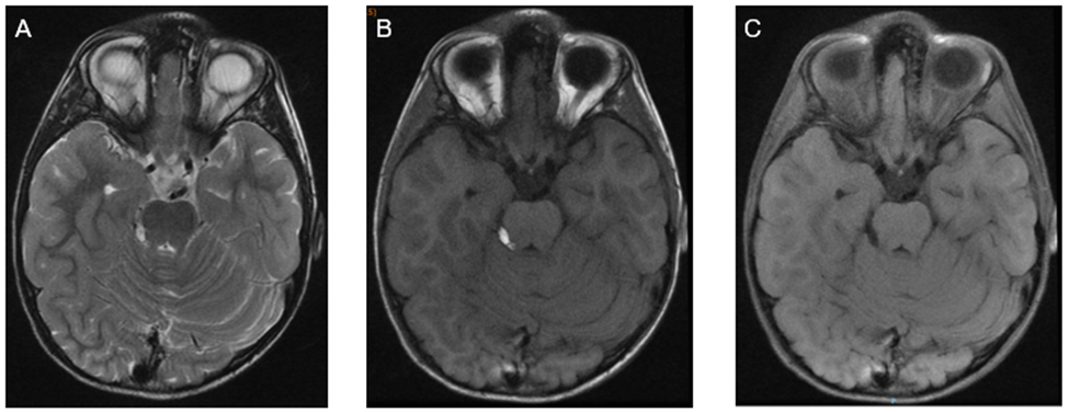
Figure 2: Brain MRI A, B, C axial slices, The images demonstrate a homogeneously oval-shaped, fat-containing suprasellar hypothalamic lesion measuring 11x 5 x 7 mm in the anterior-posterior, transverse, and craniocaudal dimensions, respectively, on the left side. The lesion is hyper intense in T1-weighted images, intermediate to hyperintense in T2-weighted images, and suppressed on fat-saturated images. No internal septation, nodularity, or calcifications are noted. lipomatous lesion in right quadrigeminal cistern 11 mm.
In the dorsal MRI, at the mid-dorsal level, presented a visible ependymal duct, greater degree of dilation, hydrosyringomyelic cavity located between T8-T10. Correctly positioned conus medullaris. Also present disc hypoplasia with congenital fusion T6-T7 and T8-T9 with remaining disc spaces with a more preserved appearance. Absence of signs of instability. No evidence of compressive pathology (Figure 3).
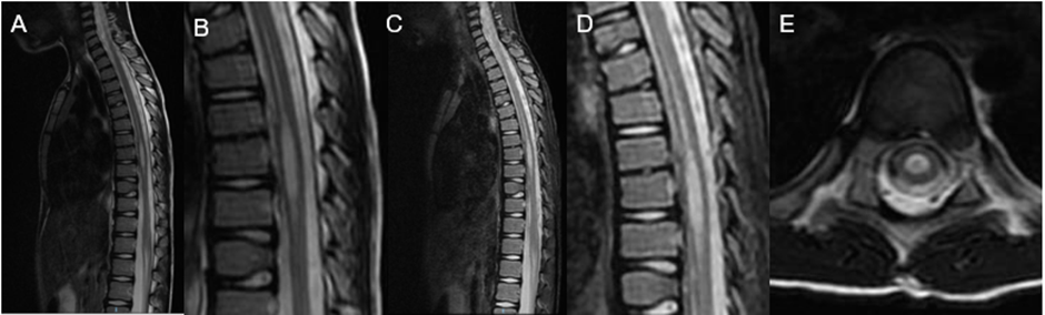
Figure 3: A, B, C, D, sagittal slices T2, E axial T2. Centromedullary syringomyelic cavity from T8 to T11, 4 x 0.4 cm
He has been referred to the rehabilitation department for presenting dorsolumbar kyphotic attitude. With the well-conducted sessions such as daily life activities and sports, he has achieved motor development identical to children of his age, fulfilling items according to his age. Although it is worth highlighting laxity in both hands, difficulty with strength and fine psychomotor skills in the hands but without signs of motor dysfunction.
In the various follow-up consultations in the Pediatrics department, normal psychomotor development was observed, and he completed schooling without problems and without dyslexia. Follow-up and evaluation with Ophthalmology showed significant astigmatism, with normal fundus and retina, normal anterior chamber.
Actually, the patient presented a good cognitive evolution, good verbal and non-verbal development, no dyslexia, great sensitivity and sense of responsibility. He is aware of his pathologies and faces them with joy, resignation, he is a fighter and an example for his family, friends and all those around him.
Below are the latest MRI images of the skull where the already known lesions are maintained without any changes in size or alterations in the different MRI weightings (Figure 4).
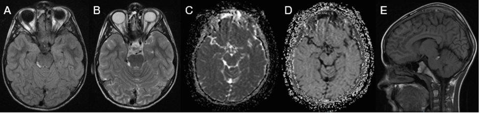
Figure 4: A, B, C, D axial slices T1, T2, ADC, eADC with lipomatous lesion in right quadrigeminal cistern. A- lipomatous lesion in anterolateral right orbit. E, T1 sagittal slice hiperintense lesion in rigth quadrigeminal cistern.
Taking into account his medical history, although he is asymptomatic, I also performed an MRI of the thoracic spine that showed normal kyphosis. Hypoplasia of the intervertebral discs T6-T7 and T8-T9. Rest of the intervertebral discs of normal height and signal intensity. Normal interfacial joints. Free conjunction foramina, of normal size and content. No rachy stenosis is observed. Centro medullary syringomyelic cavity from T8 to T11, 4 x 0.4 cm (CC x T). Regarding previous examination, a slight decrease in its maximum transverse diameter and a slight increase slight decrease in its maximum transverse diameter and a slight increase in its maximum cephalocaudal diameter stands out (previous study of 3.3 x 0.5 cm), (Figure 5). Actually, we maintain the annual follow-up with Brain and Spine MRI. Anticipate testing if the patient fell new symptoms related with the know pathologies.
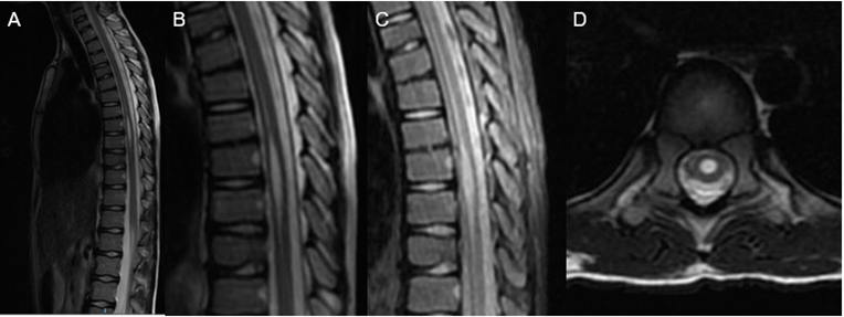
Figure 5: A, B, C, sagittal slices T2, STIR, D axial T2. Centro medullary syringomyelic cavity from T8 to T11, 4 x 0.4 cm
Intracranial lipomas (IL) are an uncommon congenital pathology that resulting from abnormal differentiation of meningeal tissue during embryonic phases of development. Most IL are small, usually pea-sized, and asymptomatic.[14] The IL are rare malformations that form due to the abnormal persistence of meninx primitiva, which is the mesenchymal tissue that the meninges arise from and differentiate into adipose tissue. These lesions are most commonly idiopathic in origin, and located mainly in the interhemispheric fissure. [15, 16] The symptoms are dependent on the location of the tumors intracranially and how they affect the surrounding brain parenchyma or neurovascular structures. The most common symptoms are headaches, seizures, or vertigo. Most commonly the patients are asymptomatic, and the IL are found as an incidental finding. [17] Diagnosis based on symptoms or clinical data needs to be complemented with neuroimaging studies such as computed tomography or magnetic resonance imaging. Nonetheless, the definitive diagnosis as in all pathologies is achieved exclusively with the histological study. [18,19, 20] Frequently can be identified structural differences at the level of the cell density between benign and malignant lesions. This study propose is determine the ADC role by comprehensively evaluating DWI and histologic features of a wide variety of pathology proven lesions. The final goal of the study is identifying specifically the added value of ADC mapping to conventional 1.5 T MRI to distinguishing the lesions into benign and malignant. In some cases, these two techniques can avoid unnecessary surgical resection and help in management guide. The different conventional sequences are useful for lesion detection. However, the DWI helps to achieve the diagnosis, providing information on the grade and type of the tumor, it also allows monitoring the response to treatment. [21] The surgical approach, for this rare benign disease with frequently slow growing behavior, is generally contraindicated. However, the surgical management has an important role in certain cases, especially in cases with hydrocephalus, uncontrollable epileptic seizures, and bone involvement with cosmetic complications. If was detected seizures, anti-seizure medications are the first line of treatment. In all the cases is recommended maintain the follow up with MRI annually at least in the 5 years following the diagnosis and whenever any symptom related to the presence of the tumor occurs. [22] Duane retraction syndrome (DRS), also called Stilling–Turk–Duane syndrome, is a congenital disease that provoke a eye movement anomaly with variable horizontal duction deficits, palpebral fissure narrowing and globe retraction during adduction, occasionally occur with upshoot or downshoot. Represents 1-5% of all forms of strabismus. [23, 24, 25] In 80% are a sporadic entity, most commonly affecting females, though familial cases have also been noted in 5%–10% of cases. [26] DRS is also more commonly unilateral and affecting more frequently the left eye. [27, 28, 29]
The etiopathogenesis of this condition can be explained by a spectrum of mechanical, innervational, neurologic and genetic abnormalities occurring independently or which influence each other giving rise to patterns of clinical presentations along with a complex set of ocular and systemic anomalies. Until now various theories were proposed to explain the etiology of DRS, many authors related this pathology with mechanical anomalies, innervational anomalies, central nervous system anomalies or the role of genetics. [30] More recently in 1998 was demonstrated the absence of left sixth nerve in a case of unilateral DRS. The authors using high-resolution T1-weighted images on magnetic resonance imaging (MRI). This congenital absence of innervation to the muscles has been found to cause fibrotic changes in the extraocular muscles leading to abnormal motility in DRS, a concept which has evolved over the last decade to be known as congenital cranial dysinnervation disorder (CCDD). [31, 32] In this disease the developmental abnormalities of one or more cranial nerves cause congenital dysinnervation of the cranial muscles. This may be primary due to absence of normal innervation or secondary following aberrant innervations from other cranial nerves. This entity, CCDD, is nonprogressive and may also have associated bony abnormalities. [33]
DRS is a group of entities linked by dysinnervation leading to limited horizontal gaze and globe retraction in attempted abduction. Understanding these mechanisms can help understand the pathogenesis of the presenting features of this syndrome. Globe retraction in adduction, which is a characteristic feature of DRS, is found to be due to tight fibrotic muscles, sometimes with anomalous insertions. Usually, these changes have been observed in the horizontal rectus muscles, as is often noted while operating such patients. DRS is a spectrum of mechanical, innervational, neurologic and genetic abnormalities that influence each other giving rise to patterns of clinical presentations which can be categorized as described below.[34] Various systems of classification were proposed to understand the mechanism and presentation of DRS. Ahluwalia and modified Huber’s classification to include further subgroups in each category described by Huber. Depending upon the alignment in primary gaze, each Huber type was divided into A, B and C, indicating esotropia, exotropia and orthophoria, respectively, thus making it even more relevant clinically and surgically. [35, 36] Huber type I DRS is the most common form of DRS with an earlier presentation, while Huber type II is the least common presentation. Usually, patients with unilateral type I Duane syndrome have esotropia more frequently than exotropia, those with type II have exotropia and those with type III have esotropia and exotropia occurring equally common. Cases of bilateral DRS may have variable presentation depending upon the type of presentation in each eye. The clinical presentation and the common reasons for seeking medical attention in DRS patients include an abnormal head position of the child, one of the eyes appearing smaller than the other due to globe retraction, pseudoptosis in adduction or abnormal eye movements. DRS is present at birth and in early stages may appear only as an abduction deficit. The inelasticity and tightness develops, the globe retraction and limitation of motility may be more pronounced with time. [37] The intelligence of the patients is usually normal in DRS but intellectual disability has been reported in a few cases with borderline intelligence and cases of autism spectrum disorder associated with fetal thalidomide exposure or HOXA1-related syndromes. [38, 39] The management of DRS is a challenge, and surgeons worldwide have their own preferred practices. In general, the surgical plan and aim of strabismus surgery in DRS is usually based on the primary position deviation, the degree of abnormal head posture, the severity of globe retraction and overshoots, degree of limitation of ductions, forced duction testing (FDT), extent of field of binocular single vision. Many authors consider that DRS classification based on primary position deviation as esotropic, exotropic or orthotropic is said to be more relevant than Huber’s classification before planning surgery. The surgical approach could be individualized based on the amount of ocular deviation, abnormal head position, associated globe retraction and overshoots present by the patients. 30 Our patient present also syringomyelia, a neurological disorder in which a fluid-filled cyst (syrinx) forms within the spinal cord. Is a neurologic condition caused by the presence of a fluid-filled cavity within the spinal cord parenchyma or central canal. Is the development of a fluid-filled cyst within the spinal cord. The cyst, which is sometimes called a syrinx, can grow larger over time. The cavity can growth, compress and damage the spinal cord. [40, 41] The etiology of this conditions are related and include conditions that alter the physiologic cerebrospinal fluid (CSF) circulation dynamics. In most cases, it is secondary to spinal subarachnoid space obstruction. This pathology has several possible causes, many cases are associated with a Chiari malformation. [42] Although other causes of syringomyelia include spinal cord tumor, trauma, and post-traumatic or infectious adhesive arachnoiditis. [43, 44] Epidemiological data related with syringomyelia is limited, the prevalence of this pathology is 8.4/100,000 to 0.9/10,000 with ethnic and geographic variation. The age of the majority of the patients in the moment of diagnosis range between 20 to 50 years. [45,46] The syringomyelia may present with sensory symptoms such as pain and temperature insensitivity, it is more commonly found incidentally. The symptoms vary among individuals depending on where the syrinx forms, how large it is, and how long it extends. The symptoms develop slowly over time, worsen over many years, and may occur on one or both sides of the body. The most common symptoms are pain, progressive weakness, stiffness in the back, shoulders, neck, arms or legs, headaches, loss of sensitivity to pain or temperature, especially in the hands, numbness or tingling, loss of balance, loss of bowel and bladder control, sexual disfunction, and scoliosis may be the only symptom in children.[47] For the diagnosis and evaluation of this pathology the magnetic resonance image (MRI) with and without gadolinium contrast is the gold standard technique. The MRI present the relevant anatomy and allows accurate visualization of the syrinx in both sagittal and axial planes. The MRI permit found the location, size, extent of the syrinx cavity and the degree of cerebellar tonsillar ectopia. After diagnosis is recommended study syrinx progression over months or years to report the natural history of syringomyelia. The high use of the MRI for the evaluation of neck or back pain has led to increased detection of syringomyelia. [48] During diagnosis and follow up of this pathology we can also use Dynamic MRI or Cardiac Gated CINE-MRI Flow Study. Permit analyze CSF hydrodynamics non-invasively. It can diagnose CSF velocity/flow disturbance at the foramen magnum particularly in patients with <5mm>
We report a clinical case with uncommon pathologies, the first case report that combine a intracranial lipoma and a Duane retraction syndrome. IL are a congenital rare benign lesions that frequently present a slow growing behavior, the symptoms depend of the location of the lesion, and in many cases are asymptomatic. Actually the MRI are the gold standard image test diagnosis. The surgical management is infrequent and could result in high morbidity/mortality. The surgery should be considered when seizures are related with the tumor and not respond to medical treatment, hydrocephalus caused by the tumor and in those cases with aesthetic changes related to tumor growth. DRS consists of a complex set of ocular and systemic anomalies. It is caused by mechanical, genetic, embryologic and central nervous system anomalies occurring together or as an independent etiology. The management of DRS is a challenge, the surgical procedure must be individualized based on the amount of ocular deviation, abnormal head position, associated globe retraction and overshoots. The surgical treatment in syringomyelia cases is to correct the underlying causative pathophysiology, the treatment strategies are directed toward improving CSF flow dynamics. In the three pathologies is recommended maintain a long term follow up with MRI annually at least in the 5 years following the diagnosis and whenever any symptom related to the presence of the tumor occurs.
To the patient and her parents that gave consent for their clinical history and particularly the images to be used in this paper, and permit the scientific community to learn and know more about this uncommon pathologies.
For this type of study, formal consent is not required. All images are anonymized. Ethical approval is exempted based on the guidelines of the research center at our institution. All the procedures being performed were part of the routine care.
The author report no conflicts of interest.
Clearly Auctoresonline and particularly Psychology and Mental Health Care Journal is dedicated to improving health care services for individuals and populations. The editorial boards' ability to efficiently recognize and share the global importance of health literacy with a variety of stakeholders. Auctoresonline publishing platform can be used to facilitate of optimal client-based services and should be added to health care professionals' repertoire of evidence-based health care resources.

Journal of Clinical Cardiology and Cardiovascular Intervention The submission and review process was adequate. However I think that the publication total value should have been enlightened in early fases. Thank you for all.

Journal of Women Health Care and Issues By the present mail, I want to say thank to you and tour colleagues for facilitating my published article. Specially thank you for the peer review process, support from the editorial office. I appreciate positively the quality of your journal.

Journal of Clinical Research and Reports I would be very delighted to submit my testimonial regarding the reviewer board and the editorial office. The reviewer board were accurate and helpful regarding any modifications for my manuscript. And the editorial office were very helpful and supportive in contacting and monitoring with any update and offering help. It was my pleasure to contribute with your promising Journal and I am looking forward for more collaboration.

We would like to thank the Journal of Thoracic Disease and Cardiothoracic Surgery because of the services they provided us for our articles. The peer-review process was done in a very excellent time manner, and the opinions of the reviewers helped us to improve our manuscript further. The editorial office had an outstanding correspondence with us and guided us in many ways. During a hard time of the pandemic that is affecting every one of us tremendously, the editorial office helped us make everything easier for publishing scientific work. Hope for a more scientific relationship with your Journal.

The peer-review process which consisted high quality queries on the paper. I did answer six reviewers’ questions and comments before the paper was accepted. The support from the editorial office is excellent.

Journal of Neuroscience and Neurological Surgery. I had the experience of publishing a research article recently. The whole process was simple from submission to publication. The reviewers made specific and valuable recommendations and corrections that improved the quality of my publication. I strongly recommend this Journal.

Dr. Katarzyna Byczkowska My testimonial covering: "The peer review process is quick and effective. The support from the editorial office is very professional and friendly. Quality of the Clinical Cardiology and Cardiovascular Interventions is scientific and publishes ground-breaking research on cardiology that is useful for other professionals in the field.

Thank you most sincerely, with regard to the support you have given in relation to the reviewing process and the processing of my article entitled "Large Cell Neuroendocrine Carcinoma of The Prostate Gland: A Review and Update" for publication in your esteemed Journal, Journal of Cancer Research and Cellular Therapeutics". The editorial team has been very supportive.

Testimony of Journal of Clinical Otorhinolaryngology: work with your Reviews has been a educational and constructive experience. The editorial office were very helpful and supportive. It was a pleasure to contribute to your Journal.

Dr. Bernard Terkimbi Utoo, I am happy to publish my scientific work in Journal of Women Health Care and Issues (JWHCI). The manuscript submission was seamless and peer review process was top notch. I was amazed that 4 reviewers worked on the manuscript which made it a highly technical, standard and excellent quality paper. I appreciate the format and consideration for the APC as well as the speed of publication. It is my pleasure to continue with this scientific relationship with the esteem JWHCI.

This is an acknowledgment for peer reviewers, editorial board of Journal of Clinical Research and Reports. They show a lot of consideration for us as publishers for our research article “Evaluation of the different factors associated with side effects of COVID-19 vaccination on medical students, Mutah university, Al-Karak, Jordan”, in a very professional and easy way. This journal is one of outstanding medical journal.

Dear Hao Jiang, to Journal of Nutrition and Food Processing We greatly appreciate the efficient, professional and rapid processing of our paper by your team. If there is anything else we should do, please do not hesitate to let us know. On behalf of my co-authors, we would like to express our great appreciation to editor and reviewers.

As an author who has recently published in the journal "Brain and Neurological Disorders". I am delighted to provide a testimonial on the peer review process, editorial office support, and the overall quality of the journal. The peer review process at Brain and Neurological Disorders is rigorous and meticulous, ensuring that only high-quality, evidence-based research is published. The reviewers are experts in their fields, and their comments and suggestions were constructive and helped improve the quality of my manuscript. The review process was timely and efficient, with clear communication from the editorial office at each stage. The support from the editorial office was exceptional throughout the entire process. The editorial staff was responsive, professional, and always willing to help. They provided valuable guidance on formatting, structure, and ethical considerations, making the submission process seamless. Moreover, they kept me informed about the status of my manuscript and provided timely updates, which made the process less stressful. The journal Brain and Neurological Disorders is of the highest quality, with a strong focus on publishing cutting-edge research in the field of neurology. The articles published in this journal are well-researched, rigorously peer-reviewed, and written by experts in the field. The journal maintains high standards, ensuring that readers are provided with the most up-to-date and reliable information on brain and neurological disorders. In conclusion, I had a wonderful experience publishing in Brain and Neurological Disorders. The peer review process was thorough, the editorial office provided exceptional support, and the journal's quality is second to none. I would highly recommend this journal to any researcher working in the field of neurology and brain disorders.
Dear Agrippa Hilda, Journal of Neuroscience and Neurological Surgery, Editorial Coordinator, I trust this message finds you well. I want to extend my appreciation for considering my article for publication in your esteemed journal. I am pleased to provide a testimonial regarding the peer review process and the support received from your editorial office. The peer review process for my paper was carried out in a highly professional and thorough manner. The feedback and comments provided by the authors were constructive and very useful in improving the quality of the manuscript. This rigorous assessment process undoubtedly contributes to the high standards maintained by your journal.

International Journal of Clinical Case Reports and Reviews. I strongly recommend to consider submitting your work to this high-quality journal. The support and availability of the Editorial staff is outstanding and the review process was both efficient and rigorous.

Thank you very much for publishing my Research Article titled “Comparing Treatment Outcome Of Allergic Rhinitis Patients After Using Fluticasone Nasal Spray And Nasal Douching" in the Journal of Clinical Otorhinolaryngology. As Medical Professionals we are immensely benefited from study of various informative Articles and Papers published in this high quality Journal. I look forward to enriching my knowledge by regular study of the Journal and contribute my future work in the field of ENT through the Journal for use by the medical fraternity. The support from the Editorial office was excellent and very prompt. I also welcome the comments received from the readers of my Research Article.

Dear Erica Kelsey, Editorial Coordinator of Cancer Research and Cellular Therapeutics Our team is very satisfied with the processing of our paper by your journal. That was fast, efficient, rigorous, but without unnecessary complications. We appreciated the very short time between the submission of the paper and its publication on line on your site.

I am very glad to say that the peer review process is very successful and fast and support from the Editorial Office. Therefore, I would like to continue our scientific relationship for a long time. And I especially thank you for your kindly attention towards my article. Have a good day!

"We recently published an article entitled “Influence of beta-Cyclodextrins upon the Degradation of Carbofuran Derivatives under Alkaline Conditions" in the Journal of “Pesticides and Biofertilizers” to show that the cyclodextrins protect the carbamates increasing their half-life time in the presence of basic conditions This will be very helpful to understand carbofuran behaviour in the analytical, agro-environmental and food areas. We greatly appreciated the interaction with the editor and the editorial team; we were particularly well accompanied during the course of the revision process, since all various steps towards publication were short and without delay".

I would like to express my gratitude towards you process of article review and submission. I found this to be very fair and expedient. Your follow up has been excellent. I have many publications in national and international journal and your process has been one of the best so far. Keep up the great work.

We are grateful for this opportunity to provide a glowing recommendation to the Journal of Psychiatry and Psychotherapy. We found that the editorial team were very supportive, helpful, kept us abreast of timelines and over all very professional in nature. The peer review process was rigorous, efficient and constructive that really enhanced our article submission. The experience with this journal remains one of our best ever and we look forward to providing future submissions in the near future.

I am very pleased to serve as EBM of the journal, I hope many years of my experience in stem cells can help the journal from one way or another. As we know, stem cells hold great potential for regenerative medicine, which are mostly used to promote the repair response of diseased, dysfunctional or injured tissue using stem cells or their derivatives. I think Stem Cell Research and Therapeutics International is a great platform to publish and share the understanding towards the biology and translational or clinical application of stem cells.

I would like to give my testimony in the support I have got by the peer review process and to support the editorial office where they were of asset to support young author like me to be encouraged to publish their work in your respected journal and globalize and share knowledge across the globe. I really give my great gratitude to your journal and the peer review including the editorial office.

I am delighted to publish our manuscript entitled "A Perspective on Cocaine Induced Stroke - Its Mechanisms and Management" in the Journal of Neuroscience and Neurological Surgery. The peer review process, support from the editorial office, and quality of the journal are excellent. The manuscripts published are of high quality and of excellent scientific value. I recommend this journal very much to colleagues.

Dr.Tania Muñoz, My experience as researcher and author of a review article in The Journal Clinical Cardiology and Interventions has been very enriching and stimulating. The editorial team is excellent, performs its work with absolute responsibility and delivery. They are proactive, dynamic and receptive to all proposals. Supporting at all times the vast universe of authors who choose them as an option for publication. The team of review specialists, members of the editorial board, are brilliant professionals, with remarkable performance in medical research and scientific methodology. Together they form a frontline team that consolidates the JCCI as a magnificent option for the publication and review of high-level medical articles and broad collective interest. I am honored to be able to share my review article and open to receive all your comments.

“The peer review process of JPMHC is quick and effective. Authors are benefited by good and professional reviewers with huge experience in the field of psychology and mental health. The support from the editorial office is very professional. People to contact to are friendly and happy to help and assist any query authors might have. Quality of the Journal is scientific and publishes ground-breaking research on mental health that is useful for other professionals in the field”.

Dear editorial department: On behalf of our team, I hereby certify the reliability and superiority of the International Journal of Clinical Case Reports and Reviews in the peer review process, editorial support, and journal quality. Firstly, the peer review process of the International Journal of Clinical Case Reports and Reviews is rigorous, fair, transparent, fast, and of high quality. The editorial department invites experts from relevant fields as anonymous reviewers to review all submitted manuscripts. These experts have rich academic backgrounds and experience, and can accurately evaluate the academic quality, originality, and suitability of manuscripts. The editorial department is committed to ensuring the rigor of the peer review process, while also making every effort to ensure a fast review cycle to meet the needs of authors and the academic community. Secondly, the editorial team of the International Journal of Clinical Case Reports and Reviews is composed of a group of senior scholars and professionals with rich experience and professional knowledge in related fields. The editorial department is committed to assisting authors in improving their manuscripts, ensuring their academic accuracy, clarity, and completeness. Editors actively collaborate with authors, providing useful suggestions and feedback to promote the improvement and development of the manuscript. We believe that the support of the editorial department is one of the key factors in ensuring the quality of the journal. Finally, the International Journal of Clinical Case Reports and Reviews is renowned for its high- quality articles and strict academic standards. The editorial department is committed to publishing innovative and academically valuable research results to promote the development and progress of related fields. The International Journal of Clinical Case Reports and Reviews is reasonably priced and ensures excellent service and quality ratio, allowing authors to obtain high-level academic publishing opportunities in an affordable manner. I hereby solemnly declare that the International Journal of Clinical Case Reports and Reviews has a high level of credibility and superiority in terms of peer review process, editorial support, reasonable fees, and journal quality. Sincerely, Rui Tao.

Clinical Cardiology and Cardiovascular Interventions I testity the covering of the peer review process, support from the editorial office, and quality of the journal.

Clinical Cardiology and Cardiovascular Interventions, we deeply appreciate the interest shown in our work and its publication. It has been a true pleasure to collaborate with you. The peer review process, as well as the support provided by the editorial office, have been exceptional, and the quality of the journal is very high, which was a determining factor in our decision to publish with you.

The peer reviewers process is quick and effective, the supports from editorial office is excellent, the quality of journal is high. I would like to collabroate with Internatioanl journal of Clinical Case Reports and Reviews journal clinically in the future time.

Clinical Cardiology and Cardiovascular Interventions, I would like to express my sincerest gratitude for the trust placed in our team for the publication in your journal. It has been a true pleasure to collaborate with you on this project. I am pleased to inform you that both the peer review process and the attention from the editorial coordination have been excellent. Your team has worked with dedication and professionalism to ensure that your publication meets the highest standards of quality. We are confident that this collaboration will result in mutual success, and we are eager to see the fruits of this shared effort.

Dear Dr. Jessica Magne, Editorial Coordinator 0f Clinical Cardiology and Cardiovascular Interventions, I hope this message finds you well. I want to express my utmost gratitude for your excellent work and for the dedication and speed in the publication process of my article titled "Navigating Innovation: Qualitative Insights on Using Technology for Health Education in Acute Coronary Syndrome Patients." I am very satisfied with the peer review process, the support from the editorial office, and the quality of the journal. I hope we can maintain our scientific relationship in the long term.

Dear Monica Gissare, - Editorial Coordinator of Nutrition and Food Processing. ¨My testimony with you is truly professional, with a positive response regarding the follow-up of the article and its review, you took into account my qualities and the importance of the topic¨.

Dear Dr. Jessica Magne, Editorial Coordinator 0f Clinical Cardiology and Cardiovascular Interventions, The review process for the article “The Handling of Anti-aggregants and Anticoagulants in the Oncologic Heart Patient Submitted to Surgery” was extremely rigorous and detailed. From the initial submission to the final acceptance, the editorial team at the “Journal of Clinical Cardiology and Cardiovascular Interventions” demonstrated a high level of professionalism and dedication. The reviewers provided constructive and detailed feedback, which was essential for improving the quality of our work. Communication was always clear and efficient, ensuring that all our questions were promptly addressed. The quality of the “Journal of Clinical Cardiology and Cardiovascular Interventions” is undeniable. It is a peer-reviewed, open-access publication dedicated exclusively to disseminating high-quality research in the field of clinical cardiology and cardiovascular interventions. The journal's impact factor is currently under evaluation, and it is indexed in reputable databases, which further reinforces its credibility and relevance in the scientific field. I highly recommend this journal to researchers looking for a reputable platform to publish their studies.

Dear Editorial Coordinator of the Journal of Nutrition and Food Processing! "I would like to thank the Journal of Nutrition and Food Processing for including and publishing my article. The peer review process was very quick, movement and precise. The Editorial Board has done an extremely conscientious job with much help, valuable comments and advices. I find the journal very valuable from a professional point of view, thank you very much for allowing me to be part of it and I would like to participate in the future!”

Dealing with The Journal of Neurology and Neurological Surgery was very smooth and comprehensive. The office staff took time to address my needs and the response from editors and the office was prompt and fair. I certainly hope to publish with this journal again.Their professionalism is apparent and more than satisfactory. Susan Weiner

My Testimonial Covering as fellowing: Lin-Show Chin. The peer reviewers process is quick and effective, the supports from editorial office is excellent, the quality of journal is high. I would like to collabroate with Internatioanl journal of Clinical Case Reports and Reviews.

My experience publishing in Psychology and Mental Health Care was exceptional. The peer review process was rigorous and constructive, with reviewers providing valuable insights that helped enhance the quality of our work. The editorial team was highly supportive and responsive, making the submission process smooth and efficient. The journal's commitment to high standards and academic rigor makes it a respected platform for quality research. I am grateful for the opportunity to publish in such a reputable journal.

My experience publishing in International Journal of Clinical Case Reports and Reviews was exceptional. I Come forth to Provide a Testimonial Covering the Peer Review Process and the editorial office for the Professional and Impartial Evaluation of the Manuscript.

I would like to offer my testimony in the support. I have received through the peer review process and support the editorial office where they are to support young authors like me, encourage them to publish their work in your esteemed journals, and globalize and share knowledge globally. I really appreciate your journal, peer review, and editorial office.

Dear Agrippa Hilda- Editorial Coordinator of Journal of Neuroscience and Neurological Surgery, "The peer review process was very quick and of high quality, which can also be seen in the articles in the journal. The collaboration with the editorial office was very good."

I would like to express my sincere gratitude for the support and efficiency provided by the editorial office throughout the publication process of my article, “Delayed Vulvar Metastases from Rectal Carcinoma: A Case Report.” I greatly appreciate the assistance and guidance I received from your team, which made the entire process smooth and efficient. The peer review process was thorough and constructive, contributing to the overall quality of the final article. I am very grateful for the high level of professionalism and commitment shown by the editorial staff, and I look forward to maintaining a long-term collaboration with the International Journal of Clinical Case Reports and Reviews.

To Dear Erin Aust, I would like to express my heartfelt appreciation for the opportunity to have my work published in this esteemed journal. The entire publication process was smooth and well-organized, and I am extremely satisfied with the final result. The Editorial Team demonstrated the utmost professionalism, providing prompt and insightful feedback throughout the review process. Their clear communication and constructive suggestions were invaluable in enhancing my manuscript, and their meticulous attention to detail and dedication to quality are truly commendable. Additionally, the support from the Editorial Office was exceptional. From the initial submission to the final publication, I was guided through every step of the process with great care and professionalism. The team's responsiveness and assistance made the entire experience both easy and stress-free. I am also deeply impressed by the quality and reputation of the journal. It is an honor to have my research featured in such a respected publication, and I am confident that it will make a meaningful contribution to the field.

"I am grateful for the opportunity of contributing to [International Journal of Clinical Case Reports and Reviews] and for the rigorous review process that enhances the quality of research published in your esteemed journal. I sincerely appreciate the time and effort of your team who have dedicatedly helped me in improvising changes and modifying my manuscript. The insightful comments and constructive feedback provided have been invaluable in refining and strengthening my work".

I thank the ‘Journal of Clinical Research and Reports’ for accepting this article for publication. This is a rigorously peer reviewed journal which is on all major global scientific data bases. I note the review process was prompt, thorough and professionally critical. It gave us an insight into a number of important scientific/statistical issues. The review prompted us to review the relevant literature again and look at the limitations of the study. The peer reviewers were open, clear in the instructions and the editorial team was very prompt in their communication. This journal certainly publishes quality research articles. I would recommend the journal for any future publications.

Dear Jessica Magne, with gratitude for the joint work. Fast process of receiving and processing the submitted scientific materials in “Clinical Cardiology and Cardiovascular Interventions”. High level of competence of the editors with clear and correct recommendations and ideas for enriching the article.

We found the peer review process quick and positive in its input. The support from the editorial officer has been very agile, always with the intention of improving the article and taking into account our subsequent corrections.

My article, titled 'No Way Out of the Smartphone Epidemic Without Considering the Insights of Brain Research,' has been republished in the International Journal of Clinical Case Reports and Reviews. The review process was seamless and professional, with the editors being both friendly and supportive. I am deeply grateful for their efforts.

To Dear Erin Aust – Editorial Coordinator of Journal of General Medicine and Clinical Practice! I declare that I am absolutely satisfied with your work carried out with great competence in following the manuscript during the various stages from its receipt, during the revision process to the final acceptance for publication. Thank Prof. Elvira Farina

Dear Jessica, and the super professional team of the ‘Clinical Cardiology and Cardiovascular Interventions’ I am sincerely grateful to the coordinated work of the journal team for the no problem with the submission of my manuscript: “Cardiometabolic Disorders in A Pregnant Woman with Severe Preeclampsia on the Background of Morbid Obesity (Case Report).” The review process by 5 experts was fast, and the comments were professional, which made it more specific and academic, and the process of publication and presentation of the article was excellent. I recommend that my colleagues publish articles in this journal, and I am interested in further scientific cooperation. Sincerely and best wishes, Dr. Oleg Golyanovskiy.

Dear Ashley Rosa, Editorial Coordinator of the journal - Psychology and Mental Health Care. " The process of obtaining publication of my article in the Psychology and Mental Health Journal was positive in all areas. The peer review process resulted in a number of valuable comments, the editorial process was collaborative and timely, and the quality of this journal has been quickly noticed, resulting in alternative journals contacting me to publish with them." Warm regards, Susan Anne Smith, PhD. Australian Breastfeeding Association.

Dear Jessica Magne, Editorial Coordinator, Clinical Cardiology and Cardiovascular Interventions, Auctores Publishing LLC. I appreciate the journal (JCCI) editorial office support, the entire team leads were always ready to help, not only on technical front but also on thorough process. Also, I should thank dear reviewers’ attention to detail and creative approach to teach me and bring new insights by their comments. Surely, more discussions and introduction of other hemodynamic devices would provide better prevention and management of shock states. Your efforts and dedication in presenting educational materials in this journal are commendable. Best wishes from, Farahnaz Fallahian.

Dear Maria Emerson, Editorial Coordinator, International Journal of Clinical Case Reports and Reviews, Auctores Publishing LLC. I am delighted to have published our manuscript, "Acute Colonic Pseudo-Obstruction (ACPO): A rare but serious complication following caesarean section." I want to thank the editorial team, especially Maria Emerson, for their prompt review of the manuscript, quick responses to queries, and overall support. Yours sincerely Dr. Victor Olagundoye.

Dear Ashley Rosa, Editorial Coordinator, International Journal of Clinical Case Reports and Reviews. Many thanks for publishing this manuscript after I lost confidence the editors were most helpful, more than other journals Best wishes from, Susan Anne Smith, PhD. Australian Breastfeeding Association.

Dear Agrippa Hilda, Editorial Coordinator, Journal of Neuroscience and Neurological Surgery. The entire process including article submission, review, revision, and publication was extremely easy. The journal editor was prompt and helpful, and the reviewers contributed to the quality of the paper. Thank you so much! Eric Nussbaum, MD

Dr Hala Al Shaikh This is to acknowledge that the peer review process for the article ’ A Novel Gnrh1 Gene Mutation in Four Omani Male Siblings, Presentation and Management ’ sent to the International Journal of Clinical Case Reports and Reviews was quick and smooth. The editorial office was prompt with easy communication.

Dear Erin Aust, Editorial Coordinator, Journal of General Medicine and Clinical Practice. We are pleased to share our experience with the “Journal of General Medicine and Clinical Practice”, following the successful publication of our article. The peer review process was thorough and constructive, helping to improve the clarity and quality of the manuscript. We are especially thankful to Ms. Erin Aust, the Editorial Coordinator, for her prompt communication and continuous support throughout the process. Her professionalism ensured a smooth and efficient publication experience. The journal upholds high editorial standards, and we highly recommend it to fellow researchers seeking a credible platform for their work. Best wishes By, Dr. Rakhi Mishra.
